Featured image with Krystyna Gieniec
Posted by FocalPlane, on 1 August 2025
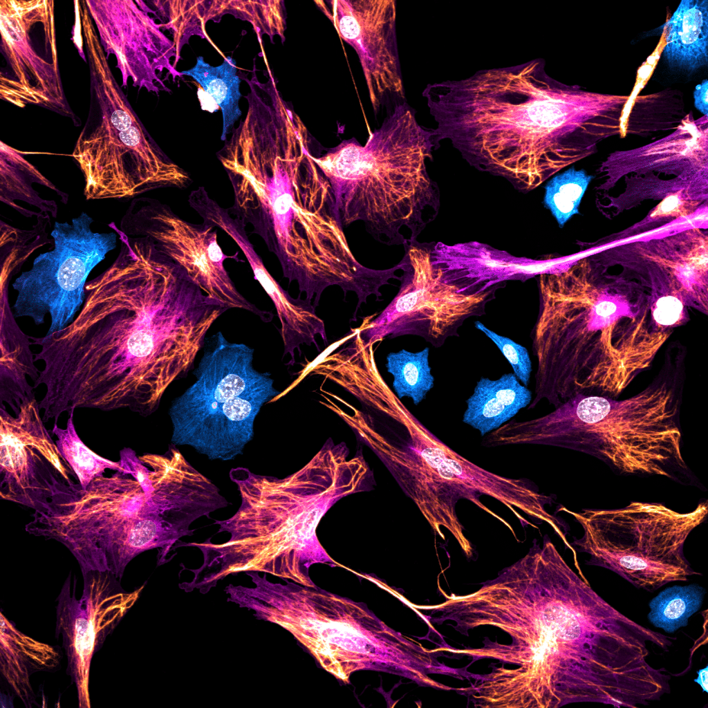
Our featured image, ‘Unexpected guests’ acquired by Krystyna Gieniec, is a 2D culture of mouse mammary fibroblasts stained for Acta2 (magenta) and Vimentin (gold), with some contaminating epithelial cells stained for pan-Cytokeratin (cyan). The image was acquired using a Leica Stellaris 8 confocal microscope.
Discover more about Krystyna’s research:
Research career so far: I have always been fascinated by cancer; the way our organs can become dysregulated and turn against themselves in such a deadly way, all in the name of evolutionary competence. I have therefore sought and designed projects in the cancer field throughout my research career, first investigating colorectal cancer during my Honours (2016) and PhD (2017-2021) degrees in the lab of A/Prof Daniel Worthley and A/Prof Susan Woods at the University of Adelaide (Australia) to then investigating breast cancer during my Postdoctoral Fellowship (2021-) in the lab of Dr Felicity Davis at the University of New South Wales Sydney (Australia). My expertise also lies in the field of fibroblast biology, particularly how cancer-associated fibroblasts contribute to the aggressive growth of tumours.
Current research: I am currently using genetically modified mouse models and complex 3D cell systems coupled with volumetric microscopy (in vitro + ex vivo + in situ) to understand how breast cancer cells and their associated fibroblasts communicate via calcium signalling to drive breast tumour growth, so that I can ultimately disrupt this signalling as a potential novel treatment strategy. In addition, I am investigating the fibroblast landscape in young vs old mice and nulliparous vs parous mice to try understand why some women are more prone to developing breast cancer as they age and in the 5 years following childbirth.
Favourite imaging technique/microscope: I consider myself extremely lucky to be part of the University of New South Wales, as they host the largest microscopy facility in Australia and I am truly spoilt by choice! My calcium imaging work requires fast imaging speeds (~500ms/z-stack) and moderate image resolution while maintaining cell viability over relatively long periods of time (~20-40 minutes), so I find myself favouring confocal microscopy and have obtained some beautiful data. However, I am now stepping into the intravital imaging space and am really looking forward to learning more about this crazy complicated (but extremely cool) technique along with multiphoton microscopy to reach deeper, healthier parts of mammary tumour tissue.
What are you most excited about in microscopy? I think there’s a lot to be excited about in microscopy as we continue to gain knowledge and improve our imaging systems, techniques and analysis pipelines! For me, I am particularly excited about new calcium biosensor development, as well as systems capable of capturing data faster with higher resolution and less phototoxicity, meaning that I’ll be able to obtain a clearer snapshot of calcium signalling in thicker sections of breast tumours. It is also equally important to know how to then analyse these increasingly complex data.
 (No Ratings Yet)