Featured image with Mathieu Preußner
Posted by FocalPlane, on 12 September 2025
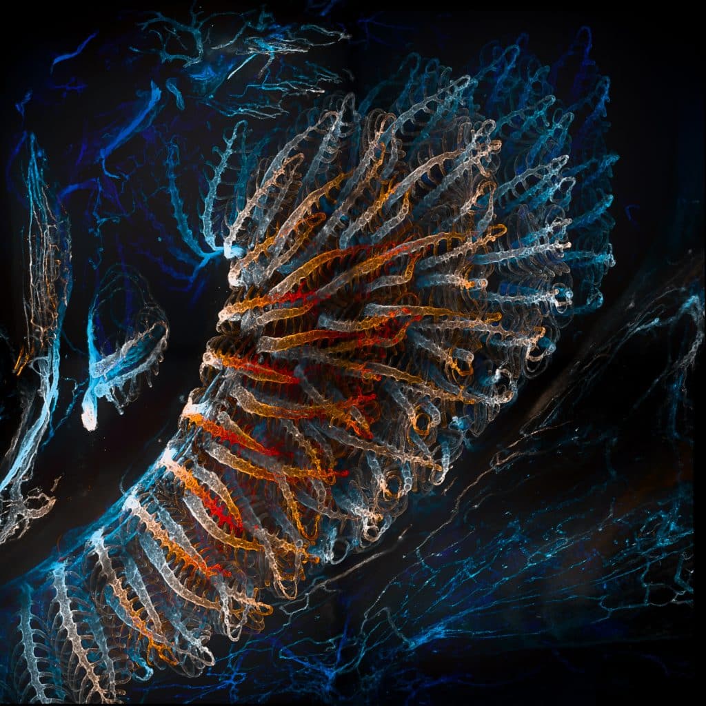
Our featured image, acquired by Mathieu Preußner, is a lateral view of the overlying gill arches in 1-month-old Danio rerio expressing endothelial kdrl:mCherry. Clarity-based tissue clearing of the sample enabled comprehensive image acquisition using a Nikon Ti spinning disk system. In ImageJ, the hyperstack was modified using a temporally colour-coded lookup table.
Discover more about Mathieu’s research.
Research career so far: I am currently in the final phase of my PhD at the lab of Virginie Lecaudey, Johan Wolfgang Goethe University, Frankfurt am Main. My scientific career began at Philipps University Marburg, where I was introduced to Drosophila developmental genetics, which sparked my intrinsic interest in developmental processes. During this time, I studied the role of an FGF receptor in myoblast fusion during flight musculature development. Later, I transitioned to biomedical sciences, seeking more translational research approaches. There, I investigated commensal bacteria-driven de novo angiogenesis in the colon of gnotobiotic mice, which is required to combat pathogen infection. I realized I truly enjoyed imaging and working with animals, which led me to pursue zebrafish for my PhD, as they are ideally suited for imaging studies.
Current research: My research initially focused on the role of Shroom3, an actin-binding protein required for apical constriction, in the kidney. However, we discovered a severe and novel phenotype in the gills of our mutants, which led us to shift the entire project toward studying this unusual and surprisingly underexplored organ. I am now investigating the temporal processes and cellular arrangements that guide gill development with a special focus on Shroom3 and its mechanobiological aspects.
Favourite imaging technique/microscope: My favourite technique has to be tissue clearing. I really enjoy exploring new structures. It’s especially fascinating to think about how these structures appear in 3D compared to conventional 2D imaging techniques. It feels like entering a new (super complex) world.
What are you most excited about in microscopy? I can’t really pick just one thing—the pace of technological advances is incredible. So many cool new opportunities are popping up. But even the ‘simple things’ like storage can be major headaches. These massive files need tons of space, and analyzing them takes serious computing power, especially with live-cell microscopy, time-lapse imaging, AI, and higher resolution—it all adds up. Still, I think with these improvements, watching an entire organism grow from a single cell will be even more mind blowing and shed light into more still unknown mechanisms.
 (No Ratings Yet)