Vote for your favourite ‘Featured image’ from 2025
Posted by FocalPlane, on 19 December 2025
Throughout 2025, we highlighted some fantastic images and researchers in our ‘Featured image’ series. Here is your chance to vote for your favourite*. The winning image will receive £100. You can vote in the poll at the end of the post.
* images that were finalists in previous image competitions are excluded from this vote.
Voting closes on 31 December 2025
We’re currently looking for images for our 2026 series, which will be kicking off in the new year. Please send us your nominations, which can be self-nominations!
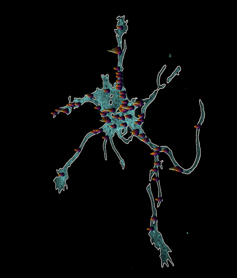
1. Exocytic hotspots
This featured image, from Ellen O’Shaughnessy, shows the locations of exocytic hotspots in a developing cortical neuron. Ellen O’Shaughnessy, Stephanie Gupton and colleagues developed pHusion, a toolkit for analysis of exocytosis that was published in Journal of Cell Science’s special issue on ‘Imaging Cell Architecture and Dynamics‘.
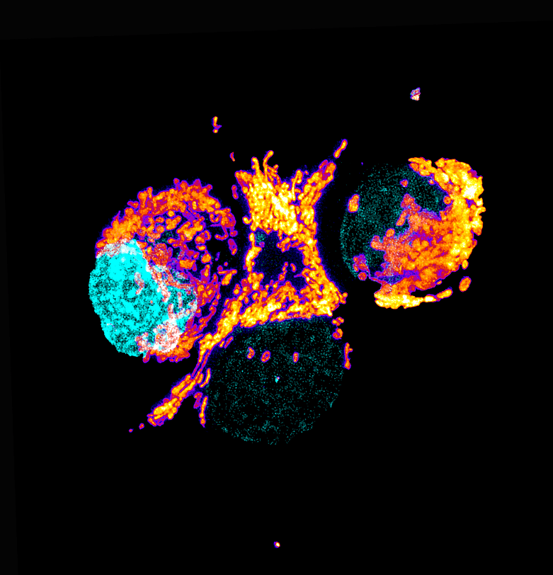
2. Mitochondria in PDAC cells
This featured image, acquired by Anthony Dornan and Fara van der Schans, showcases the perinuclear distribution of mitochondria in the highly respiratory human pancreatic ductal adenocarcinoma (PDAC) tumour cells (Panc-1) stained with the mitochondrial stain TMRE (thermal LUT), the mitochondria are associated with the nuclei, stained with DAPI (cyan).
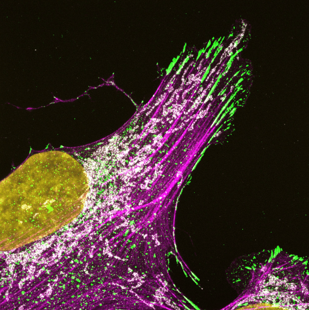
3. Paxillin, mitchondria and actin
This featured image, acquired by Omkar Joshi, showcases a U2OS cell immunostained for paxillin (green), TOMM20 (mitochondria, white), and F-actin (labelled using SiR-Actin, magenta), with DAPI (yellow) marking the nucleus.
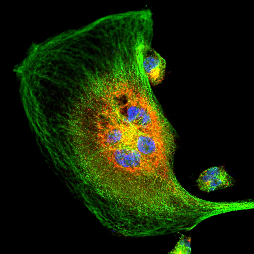
4. A dividing MDA-MB-231 cancer cell
This featured image, acquired by Subhajit Karmakar, depicts a dividing MDA-MB-231 cancer cell that has been stained to visualise the localisation of two key proteins: CK2, a highly oncogenic kinase, and an E3 ligase, a tumour suppressor.
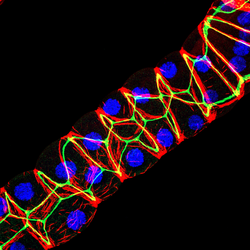
5. Malpighian tubules
This featured image, acquired by Saurabh Chand Sagar, presents a laser scanning confocal projection of the Malpighian tubules (MTs) from third instar wandering-stage (118 ± 2 hours post-eclosion) wild-type Drosophila melanogaster larvae.
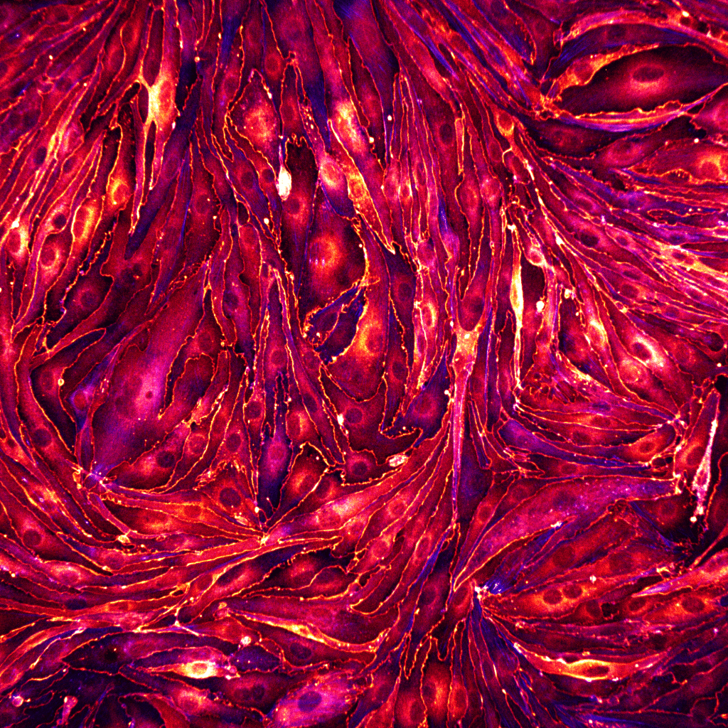
6. Endothelial cells
This featured image, acquired by Rohit Nautiyal, shows a monolayer of brain endothelial cells seeded on a glass substrate coated with fibronectin. The cells were fixed and stained for ZO-1 (LUT- Red Hot) to visualize the tight junctions and actin (LUT-blue).
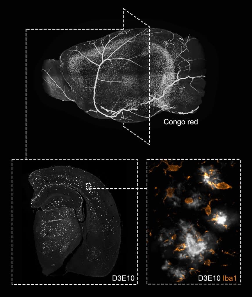
7. Mouse brain
This featured image, acquired by Andrew Octavian Sasmita, depicts a single brain hemisphere of the 5xFAD mouse model of Alzheimer’s disease (AD) at different spatial scales.
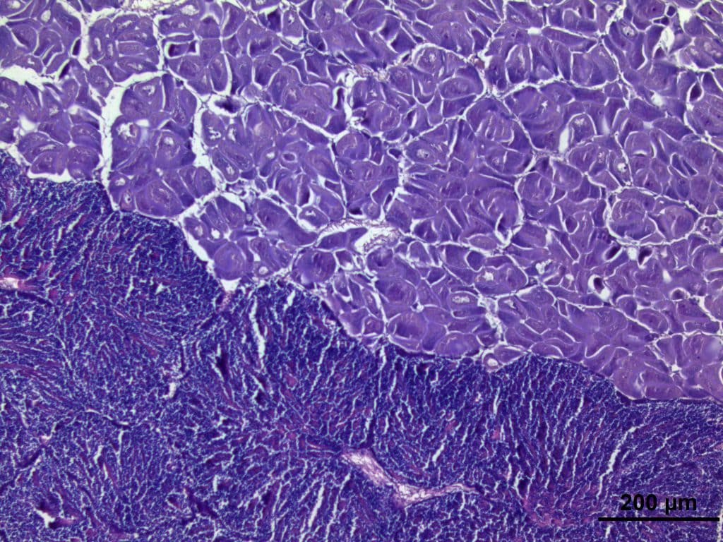
8. Scallop
This featured image, acquired by Rosa L. Salgado García, shows a histological section of female (above) and male (below) gonads at an advanced maturation stage of the simultaneous hermaphroditic scallop Nodipecten subnodosus.
9. Nuclear IRF9
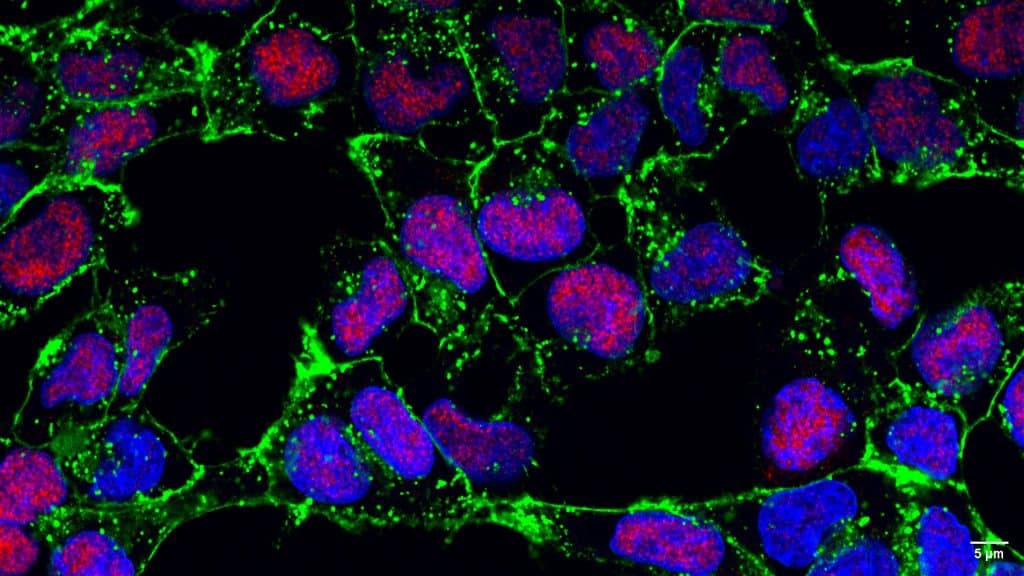
This featured image, acquired by Seth Agyei Domfeh, shows the nuclear localisation of interferon regulatory factor 9 (IRF9) in human hepatoma (HepG2) cells stimulated with interferon α (IFN-α).
10. Nematode
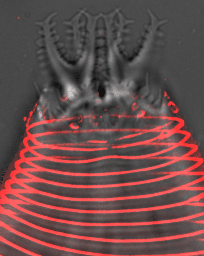
This featured image, acquired by Kaiden Power, shows the head and probolae of an Acrobeles complexus nematode. The cuticle furrows (annuli) and cilia pores are stained with Concanavalin A (red).

11. Cerebral organoid
This featured image, acquired by Aswathy G Krishnan, depicts a neural rosette of a cerebral organoid derived from human induced pluripotent stem cells (hiPSC).
Vote here by 31 December 2025:
Thank you for voting
 (21 votes, average: 1.00 out of 1)
Image 5. Magnificent visualization.
5
Image 5, malpighian tubules
Vivid multicolour depiction of Malpighian tubules
Image is quite clear and informative
Very magnificent
6
Magnificent
Image 5 a proper visualization
Brilliant
5
The most crystal clear.
Image 5:Malpighian tubules
Fantatic
3
Vote for image 3
3
3rd image by Omkar Joshi
Voted for image no 3
Excellent
5