Featured image with Ivan Radin
Posted by FocalPlane, on 16 January 2026
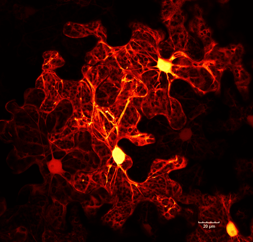
Our featured image, acquired by Ivan Radin, shows a few epidermal cells of tobacco (Nicotiana benthamiana) leaf, expressing cytosolic GFP. There is a significant variability in expression levels between cells. The brightest region is the nucleus, which is surrounded by a large central vacuole. The cytosol is restricted to the cell periphery. Transvacuolar strands connect the area around the nucleus with other parts of the cell periphery.
The image was captured on an Olympus/Evident Confocal FV3000 with HyD detectors using 60x/1.2W objective. A small section of the live leaf was mounted in water. The final image is a cropped region of a larger 3-by-3-stitched Z-stack image that was deconvolved. The fall (black to red to orange to yellow to white) look-up table has been applied.
Discover more about Ivan’s research.
Research career so far: I completed my PhD at Technische Universität Dresden, where I worked on the mitochondrial copper chaperones in plants and yeast. Then, I did my postdoc in the lab of Elizabeth Haswell at Washington University in St. Louis, where I focused on the functions of PIEZO mechanosensitive ion channels in the green lineage. In the summer of 2023, I started my lab at the Department of Plant and Microbial Biology, University of Minnesota.
Current research: We are trying to better understand the evolution of organellar mechanosensing in the green lineage (plants and green algae). More specifically, we are investigating how vacuoles and chloroplasts in Arabidopsis, the moss Physcomitrium patens, and the green alga Chlamydomonas reinhardtii perceive and respond to mechanical forces.
Favourite imaging technique/microscope: Point scanning confocal. Love doing live, multicoloured, high-resolution imaging to observe changes in the morphology of different organelles. I am biased, but I love my Olympus FV3000.
What are you most excited about in microscopy?
I am really looking forward to further improvements in near-infrared and infrared imaging, as this is gentler on live samples and will expand our repertoire of available colours for multicolour imaging.
 (No Ratings Yet)