Imaging spotlight: visualising synaptic vesicle fusion by cryo-ET
Posted by FocalPlane, on 21 January 2026
In this paper highlight from Jana Kroll and colleagues, we hear about their research combing optogenetic stimulation with plunge freezing and cryo-electron tomography to study synaptic vesicle fusion.
What are the key results from your paper?
In our paper, we developed a workflow to visualize synaptic vesicle fusion in neuronal synapses under near-native conditions. By combining optogenetic stimulation with plunge freezing and cryo-electron tomography, we captured neuronal synapses only 2-3 milliseconds after an action potential.
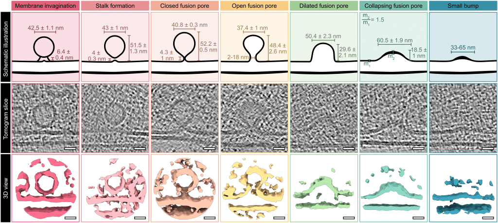
Within these synapses, we observed all putative vesicle fusion intermediates – from fusion pore formation through pore opening, collapse, and incorporation into the cell membrane – indicating that the entire fusion process takes only a few milliseconds.
Our images further revealed that membrane fusion is already initiated through a stalk-like connection when the synaptic vesicle is still several nanometers away from the cell membrane. This finding challenges the long-standing idea that synaptic vesicles reach a tightly docked state, characterized by broad membrane contact, prior to fusion.
In addition, we observed that the majority of fusing vesicles are physically connected to at least one neighboring synaptic vesicle by thin filamentous tethers. We suggest that these connectors may facilitate the fast resupply of vesicles after fusion, which is particularly relevant in periods of high synaptic activity.
Which imaging techniques have you used in this research?
We used timed cryo-electron tomography for the quantitative, morphometric characterization of synaptic vesicle fusion in neurons. To verify synaptic activity, we performed cryo-confocal microscopy of the genetically encoded glutamate sensor iGluSnFR3.
What makes this analysis special is that iGluSnFR3 is a biosensor with very fast on- and off-kinetics, typically applied in live fluorescence microscopy. By synchronizing neuronal stimulation with rapid cryo-fixation, we were able to preserve the glutamate-bound, highly fluorescent state of iGluSnFR3, making it compatible with cryo-confocal imaging. For a subset of our experiments, we combined iGluSnFR3 cryo-confocal imaging with cryo-electron tomography in a correlative cryo-CLEM approach, allowing us to directly link high fluorescence intensity to ultrastructural changes during synaptic activity.
Are there technical tips or tricks that you can share and are you open to researchers contacting you for collaboration on the methodology?
Generally, cryo-electron tomography of neurons is technically challenging because neurons are more sensitive than many other cell types. For example, we experienced that on-grid neuronal cultures would die from one day to the next without any obvious reason. After a few attempts, and often without changing much in the setup, the same cultures survived again. The most important lesson we learned is that persistence eventually pays off, especially when working with complex experimental systems.
We are happy to share our experiences with researchers and are open for collaborations. To our knowledge, we are among the first labs with an optogenetics-compatible plunge freezer. Our setup is optimized for time-resolved cryo-electron microscopy, enabling experiments with temporal resolution in the millisecond range.
Are there any advances in imaging or image analysis that would help your research?
When we started this project roughly five years ago, thinning of cells via focused ion beam (FIB) milling was not yet established in our facility. As a result, we were limited to regions thin enough for direct cryo-electron tomography (typically below 500 nm). With FIB milling, we could have selected regions for data acquisition primarily based on the fluorescence intensity of our used glutamate sensor. The most recent cryo-FIB instruments additionally contain an integrated fluorescence microscope, which can be used to select regions for thinning with at least micrometer-scale precision.
 (No Ratings Yet)
Get involved
Create an account or log in to post your story on FocalPlane.
More posts like this

Filter by
- NewsApply
- DiscussionsApply
- How toApply
- ToolsApply
- Case studiesApply
- InterviewsApply
- JobsApply
- EducationApply
- Blog seriesApply
- Asian Microscopists ..and Cell BiologistsApply
- AIC at HHMI JaneliaApply
- Deep Learning for Bi..o-image analysisApply
- GloBIAS – updates fr..om the communityApply
- Volume EMApply
- Latin American Micro..scopistsApply
- Bio-image Analysis w..ith NapariApply
- Imaging with…Apply
- Towards Global Acces..sApply
- Latin America Bioima..gingApply
- From Zero to Qupath ..HeroApply
- Highlights from Euro..-BioImagingApply
- LSFM seriesApply
- DIY MicroscopyApply
- View all