Featured image with Emily Oren
Posted by FocalPlane, on 10 April 2026
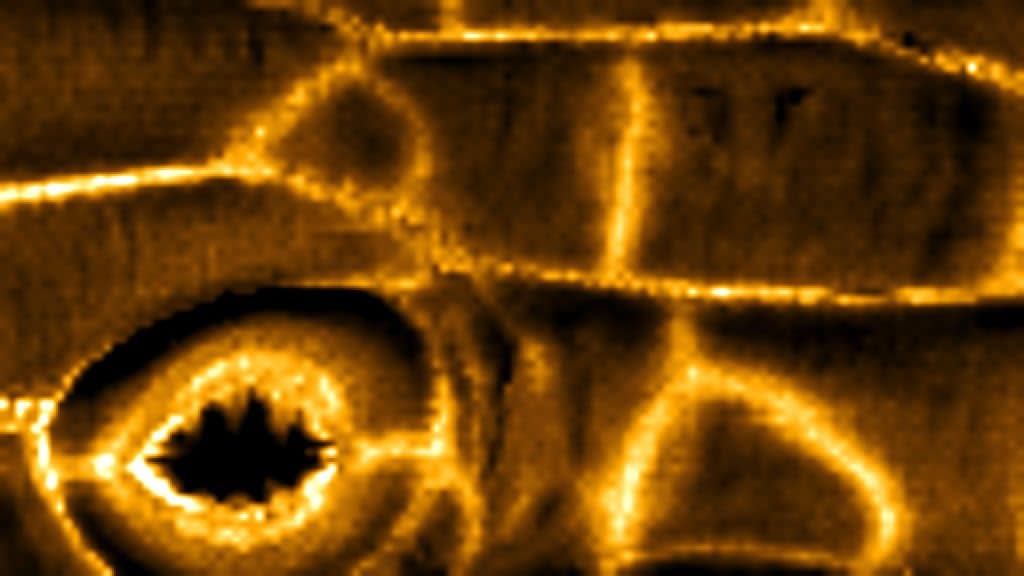
Our featured image, acquired by Emily Oren, shows the surface stiffness of an Arabidopsis thaliana hypocotyl. It was acquired using a Bruker Biowizard 5 Atomic Force Microscope (AFM) and is of plasmolysed epidermal cells on a live seedling. AFM is a mechanical imaging technique that uses a nanoscale needle to probe the surface of a sample and measure its local Young’s Modulus. In this image, brighter colours correspond with a higher Young’s Modulus. The stiff anticlinal cell walls are clearly visible, as well as the distinctive stiffness pattern of the guard cells surrounding the stomatal pore. This image was processed using JPKSPM Data Processing and Gwyddion.
Discover more about Emily’s research.
Research career so far: I completed my undergraduate degree in 2021 in Molecular, Cell, and Developmental Biology at the University of California Los Angeles (UCLA). After graduating, I continued on at UCLA as a lab technician in Dr Siobhan Braybrook’s group where I began my journey in plant developmental biology. I am currently working towards my PhD in the group of Dr Sarah Robinson, focusing on the biomechanics of plant development.
Current research: In my PhD project, I am investigating the regulatory relationships between three factors known to impact plant growth: hormonal signalling, mechanical feedback, and cell division. While all three are known to contribute to growth regulation, how each feeds back on the others is currently unclear. To begin to unravel this complex process, I study cell division in Arabidopsis hypocotyls, an organ with very few endogenous divisions, and use inducible lines that increase cell division frequency. To investigate mechanical properties, I use Atomic Force Microscopy (AFM) and an Automated Confocal Micro Extensometer (ACME). AFM allows for cell wall scale measurement of stiffness, while ACME operates at the whole organ scale. I am also using confocal microscopy, freeze-fracture cryoSEM, and single cell transcriptomics to fully investigate this system.
Favourite imaging technique/microscope: My favourite imaging technique is AFM. It’s a challenging method, but that just makes it more satisfying to get a good scan. I love the simultaneous simplicity and complexity of mechanical imaging, as well as the unique questions it allows us to investigate.
What are you most excited about in microscopy? I am most excited about the collaborations that can bring new and exciting imaging methods into our toolkit for investigating developmental biology. Working with physicists, chemists, engineers, and more has resulted in so many amazing techniques to investigate living systems with, and I can’t wait to see (and contribute to) what comes next!
 (No Ratings Yet)