

Imaging spotlight: VLab4Mic
Posted by FocalPlane, on 7 July 2026
In this preprint highlight, Damián Martínez, Bruno M. Saraiva, Tayla Shakespeare, Mario Del Rosario and Ricardo Henriques introduce VLab4Mic, an open-source simulation platform that allows researchers to optimise microscopy experiments before performing them.
Can you briefly describe what VLab4Mic does and explain why you developed the platform?
VLab4Mic is an open-source platform that lets researchers test microscopy experiments before performing them. It predicts whether biologically meaningful structural differences are likely to remain experimentally distinguishable after labelling and imaging.
Rather than asking how a structure will look under the microscope, VLab4Mic asks whether a biological hypothesis is experimentally resolvable.
We developed VLab4Mic based on questions we often face when starting or optimising protocols such as: “Will conventional microscopy be enough to resolve my structures?” or “Are two structures different enough to be seen with super-resolution?”, “How can I test if uneven labelling introduce bias?”, for instance.
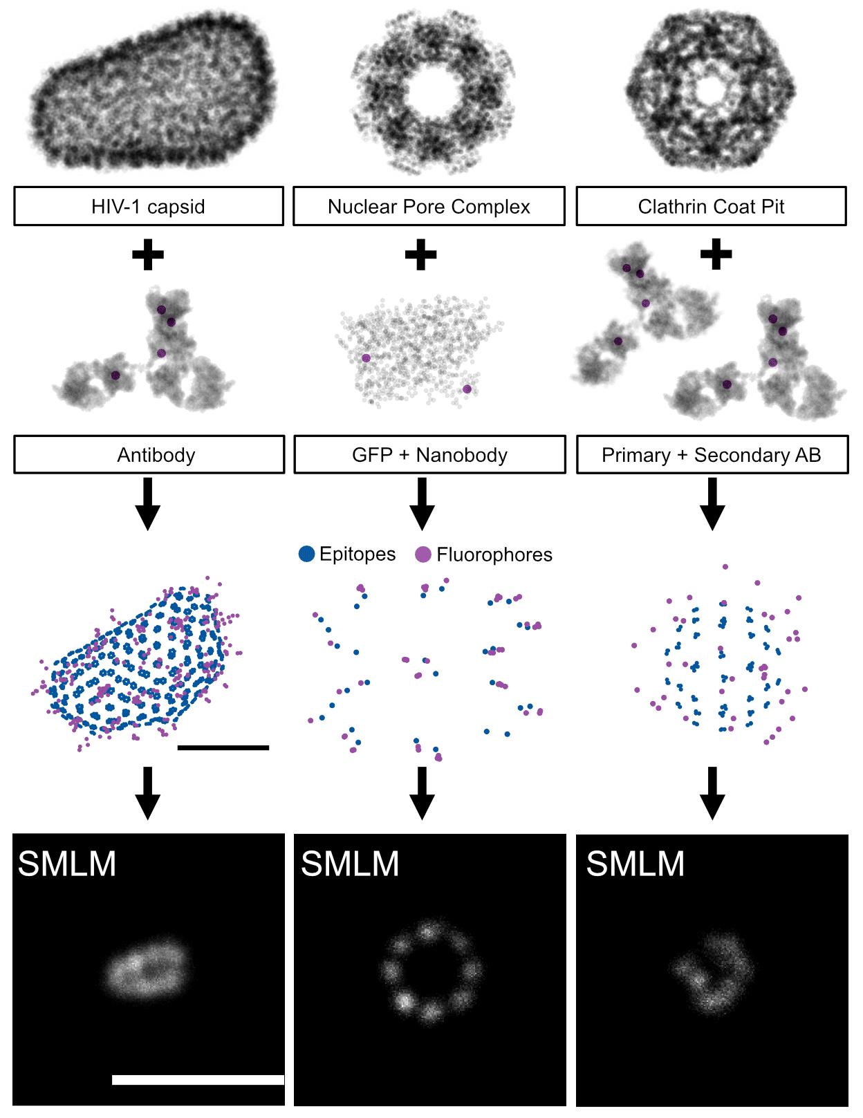
Recovering meaningful features from images is highly dependent on each step of the experimental design. For instance, when choosing a labelling strategy, a researcher’s choice on using a primary and secondary antibody, nanobodies or a fusion protein will depend on whether a labelling strategy is appropriate with the experiment and available resources. Each option carries a level of on how faithfully the structure can be labelled and if the labelling itself creates an extra layer of bias. Experiments, probes and access to imaging modalities vary across laboratories; within the same laboratory this variability means that the choice is often subjected to trial and error, which is both costly and time consuming.
VLab4Mic models the imaging experiment from the structure to the analysis. We leverage atomic models available for both the macromolecular assemblies and the probes used to target them to model the stochastic nature of sample staining. Subsequent imaging simulation and analysis in VLab4Mic enable researchers to answer two important questions: how will the same sample would look under different imaging modalities? and how will sample variation look under the same modality? For cases where high variability at the sample preparation is expected, the central challenge is therefore not simply achieving high resolution but to be able to interrogate if two functionally distinct structural states are likely to remain experimentally distinguishable under the tools available to us.
What have you modelled so far using VLab4Mic and are there any limitations where the platform might struggle?
The current limitation is that VLab4Mic relies on structural models (PDB/CIF). If a molecular assembly has no structural information available, we first need to build an approximate model, as we did for clathrin.
We first validated VLab4Mic by replicating imaging experiments for the Nuclear Pore Complex, a well characterised complex commonly used as a standard in super-resolution microscopy. Available data for the NPC meant we had one example of the same underlying structure seen in different imaging systems experimentally. With VLab4Mic, it is possible to mimic those experiments, and at the same time it allows us to extend these observations to unexplored scenarios. When we parameterised our experiments to be as close as those of the experimental data, the simulations closely matched the experimental observations between the two.
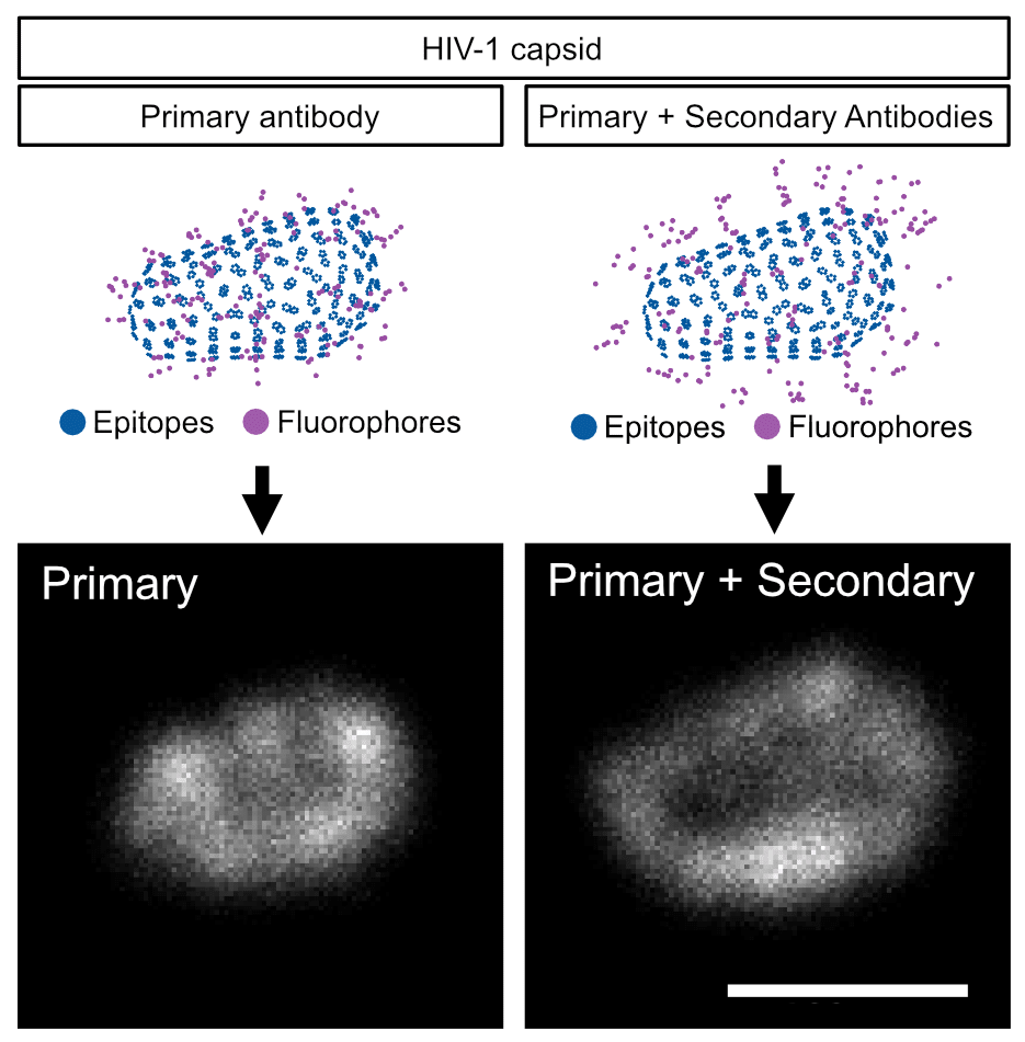
Besides NPC, we chose the HIV-1 capsid to showcase another macromolecule that has been extensively characterised, but for which multimodal examples are limited. With HIV-1 capsid we show how its appearance under the microscope varies solely by the efficiency of labelling or by choosing to label it with primary antibody alone vs with primary and secondary antibody.
Other examples of diverse biological structures include clathrin-coated pits, T4 capsids, but as PDB/CIF are the basis of structure definition, modelling of precise arrangement for epitopes is restricted to those structures with available PDB/CIF. Nonetheless, we show one way to circumvent this current limitation for the case of clathrin-coated structures (CCS). We created composite PDB/CIF models to closely match reported images of these CCS as seen through electron microscopy (EM). This means that the ultrastructure of CSS in EM is not only complementary but easy to integrate with VLab4Mic workflow.
Do you have any tips or tricks for researchers who would like to use VLab4Mic?
We encourage researchers to use VLab4Mic with our codeless Jupyter notebooks for quick and versatile exploration of its capabilities. These notebooks provide an interactive graphical interface where researchers can have immediate feedback on how their choices change the course of experiment. For example, a user can select and preview different structures before selecting what to use to label them. Once a structure is chosen, it is possible to explore how choosing different labelling strategies result in particular fluorophore distributions. This exploration extends into creating a virtual sample, which imaging modalities to use and ultimately how to integrate this exploration into a parameter sweep. We also provide tutorials and example scripts for advanced users.
What are the prospects for further development?
We envision that increasing availability and refinement of prediction of protein folding and interactions, such as with AlphaFold, will increase the catalogue of molecular assemblies whose structures are uncharacterised. Nonetheless, complementary techniques such as cryo-electron tomography (cryo-ET) could be used at the structure modelling stage at the expense of precision and accuracy at positioning epitope sites. As structural biology continues to expand through AlphaFold, cryo-EM and cryo-ET, the number of molecular assemblies that can be simulated with VLab4Mic will continue to grow.
Where can people find more information?
The preprint is available at under the name “VLab4Mic: prediction of structural resolvability in super-resolution microscopy” where detailed information can be found about case studies and examples. The code is open-source and available in GitHub (https://github.com/HenriquesLab/VLab4Mic) along manuals and tutorials on how to quickly use it. Additionally, users might refer to our dedicated website (https://vlab4mic.henriqueslab.org).
We hope VLab4Mic helps researchers spend less time wondering whether an experiment might work, and more time designing experiments they already know are capable of answering their biological question.
Get involved
Create an account or log in to post your story on FocalPlane.
More posts like this


Filter by
- NewsApply
- DiscussionsApply
- How toApply
- ToolsApply
- Case studiesApply
- InterviewsApply
- JobsApply
- EducationApply
- Blog seriesApply
- AIC at HHMI JaneliaApply
- Deep Learning for Bi..o-image analysisApply
- GloBIAS – updates fr..om the communityApply
- WAMBIAN: West Africa.. in FocusApply
- Volume EMApply
- Latin American Micro..scopistsApply
- Bio-image Analysis w..ith NapariApply
- Imaging with…Apply
- Towards Global Acces..sApply
- Latin America Bioima..gingApply
- From Zero to Qupath ..HeroApply
- Asian Microscopists ..and Cell BiologistsApply
- Highlights from Euro..-BioImagingApply
- LSFM seriesApply
- DIY MicroscopyApply
- View all