

Featured image with Luz María Fuentealba Pérez
Posted by FocalPlane, on 28 August 2026
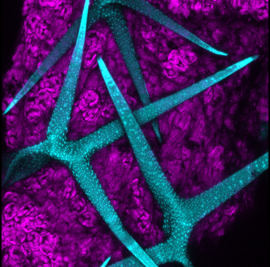
Our featured image, acquired by Luz María Fuentealba Pérez, shows the last breath of Arabidopsis thaliana, the most widely used plant model worldwide.
This organism is a small plant that has a small, fully sequenced genome and a short life cycle, which allows functional experiments to be conducted in a short amount of time. The plant was employed in an experiment conducted by researcher Catalina Saldaña from the Sakuromics lab (https://www.instagram.com/sakuromicslab/), group lead by Dr. Andrea Miyasaka. The objective of the experiment was to image the internalization of a fluorescent nanoparticle within the plant, with a view to investigating its potential as a carrier for transient genetic modification to be applied in sweet cherry buds. The image was captured immediately before discarding the untreated plant. Trichome displays autofluorescence, which was imaged on a Leica SP8 confocal microscope using a 405 nm laser, and a 633 nm laser was used to image the autofluorescence of the chlorophyll. The image is a max Z-projection across the volume, which was processed in Fiji.
Research career so far: During my undergrad studies at Universidad Austral de Chile, I did a lot of biochemistry and molecular biology (so far from microscopy!). But as a PhD student at Universidad Católica in Santiago, Chile, I used microscopy heavily throughout my project. I studied the intracellular traffic of a receptor in Lowe Syndrome, where I had to document the changes in the intracellular traffic of receptors in cell lines and i3-neurons while OCRL1, an enzyme involved in cellular membrane trafficking and actin polymerization, was not working. By the end of the PhD, I had fallen in love with microscopy. I became the Facility Manager of the UM² (Unidad de Microscopía de la Universidad Mayor). So far, I have helped users studying various topics and different kinds of samples, from classical cell cultures with multiple markers to assess the effect of different drugs on cell function, to complete Xenopus embryos that have been cleared and imaged using a lightsheet to understand how the heart develops. Moreover, I am actively involved in the local community at the newly created SoChIC (Sociedad Chilena de Imágenes Científicas) and LABI (Latin America BioImaging), while also participating in the international community at GBI (Global BioImaging) and BINA (BioImaging North America). We are very far away in Chile, so these collaborations across South America and within Chile are especially important.
Current research: As well as all the normal functions that I fulfil as facility manager, I am also involved in several projects currently running in the unit by helping researchers get the most out of our microscopes and their samples. For example, with PhD student Philippe Loustau, who works in Dr. Nicole Trefault’s group (https://www.linkedin.com/in/nicole-trefault-48443216/), I’ve been taking images of marine sponges from Chilean coastal areas and Antarctica to describe their bacterial population. I’ve also been working with Dr Cecilia Arriagada and her team (https://researchers.uss.cl/es/persons/cecilia-lorena-arriagada-momberg/) to quantify the volume of Xenopus hearts to help understand how environmental factors contribute to cardiac development and lead to congenital heart defects. Moreover, as a unit, along with Dr Charlotte Buckley, Dr Aníbal Vargas and Dr Leonardio Valdivia (https://cib.umayor.cl/microscopia/), we’re developing protocols for the optimisation of optical clearing, staining and visualisation of samples for imaging on our Lightsheet 7 microscope, in particular, to mimic histological staining in a 3D fashion.
Favourite imaging technique/microscope: My favourite imaging technique is fluorescence microscopy, in all its forms. I love how versatile it is, depending on the question you’re asking. There’s the simplicity and wonder of live imaging autofluorescence in water from a pond or moss; you never quite know what you’re going to find, and there’s something magical about seeing structures light up without any staining at all. While, combined with confocal microscopy, you can detect several markers at once, layering information to build a much richer picture of what’s happening inside a sample. And then there’s lightsheet microscopy, which is almost the opposite challenge: instead of painstaking detail, it’s about speed and scale, imaging entire organs in a fraction of the time it would take with other techniques, while still preserving beautiful resolution. What I really love is combining these approaches. Being able to move between the immediacy of live fluorescence, the precision of confocal, and the speed of lightsheet means I can adapt my imaging strategy to whatever biological question I’m chasing, rather than forcing the question to fit one single tool.
What are you most excited about in microscopy? Right now, I’m most excited about optical clearing of live organisms. Traditional optical clearing methods have opened up incredible possibilities for imaging deep into tissues, but they typically require fixation, which means you lose the ability to observe dynamic, living processes. The idea of clearing tissue in live organisms feels like it could be a real turning point for the field. It would let us watch developmental processes, disease progression, or physiological responses unfold in real time, at depths and resolutions that just aren’t possible today without sacrificing the sample. I think this could fundamentally change how we study whole-organism biology, moving us away from static snapshots and toward truly dynamic, longitudinal imaging of living systems.