

Register now for FocalPlane features… quantitative plant imaging
Posted by FocalPlane, on 23 September 2026
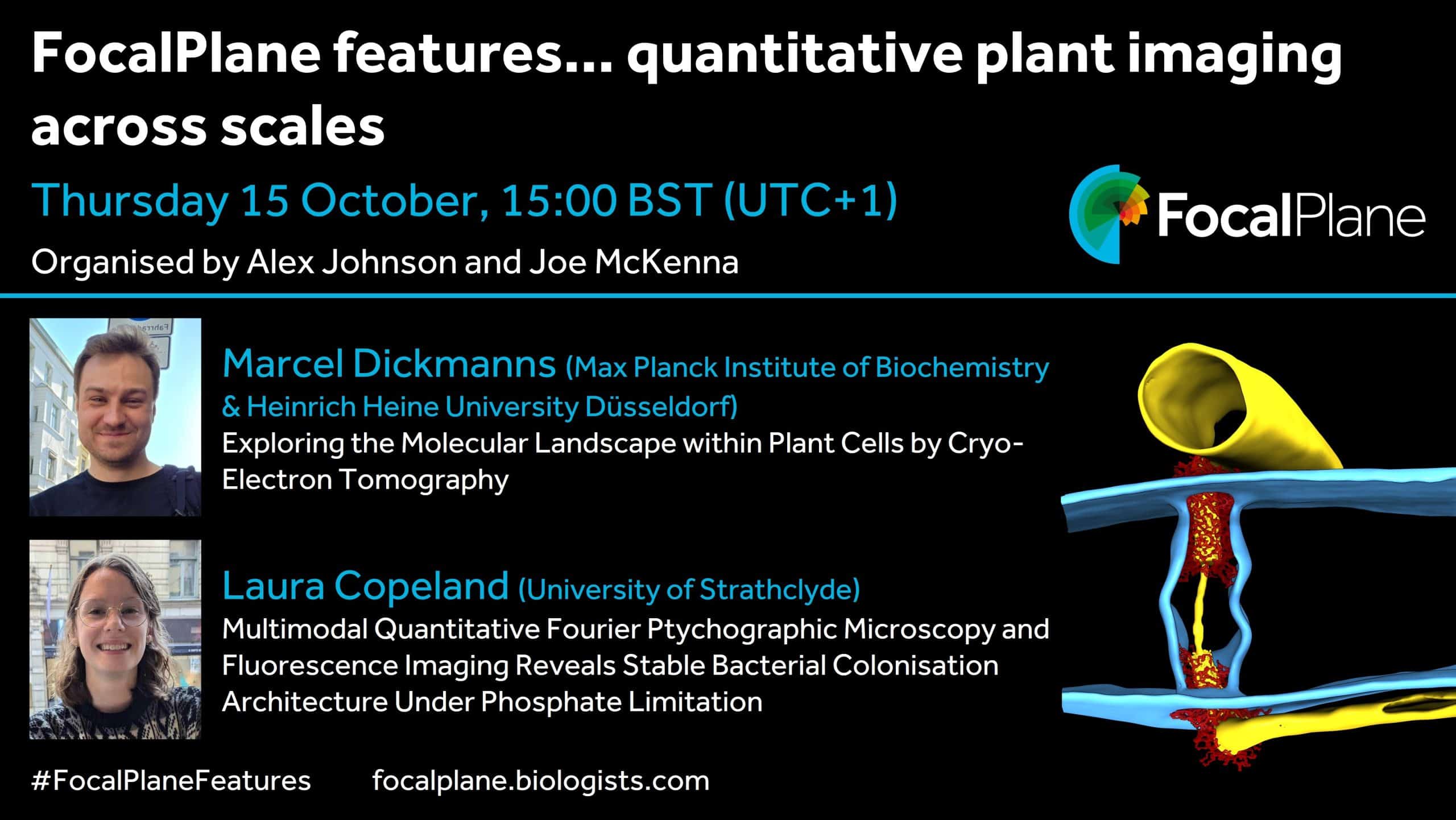
October brings the third instalment in our webinar series on quantitative plant imaging, organised by Alex Johnson and Joe McKenna. In this webinar, we’ll hear from Marcel Dickmanns on ‘Exploring the Molecular Landscape within Plant Cells by Cryo-Electron Tomography’ and Laura Copeland on ‘Multimodal Quantitative Fourier Ptychographic Microscopy and Fluorescence Imaging Reveals Stable Bacterial Colonisation Architecture Under Phosphate Limitation’. We invite you to join us on Thursday 15 October at 15:00 BST (UTC+1).
The goal of this new series is to bring together researchers working at different scales to begin understanding how we can integrate them to generate holistic understanding of plant biology from the single molecule to whole plants. We will also invite speakers from outside the plant fields to enable cross model knowledge and technical exchange. You can catch up on previous webinars on our dedicated webpage.
Our next webinar will be on Thursday 21 January 2027 at 15:00 GMT and we’d love to hear from researchers that would like to share their research with the community.
Marcel Dickmanns, Max Planck Institute of Biochemistry & Heinrich Heine University Düsseldorf
Exploring the Molecular Landscape within Plant Cells by Cryo-Electron Tomography
Cryo-electron tomography (cryo-ET) enables the visualization of macromolecules directly within their native cellular context, but its application to plants has so far been hindered by technical challenges. We have developed workflows enabling high-resolution 3D imaging of proteins, membrane interfaces and cell walls within vitrified plant tissue. Applying cryo-ET to the moss Physcomitrium patens, we determined the in situ architecture of plasmodesmata, revealing cell wall-based gating mechanisms in detail and resolving the structure of the protein assemblies scaffolding the central ER-derived desmotubule.
Integrated with complementary approaches such as mass spectrometry, fluorescence microscopy and structural bioinformatics, cryo-ET allows building mechanistic models of cellular architecture and function. By extending these workflows to other specimens, from multicellular green algae to angiosperms, we aim to facilitate broader application of in situ structural biology within the plant sciences.
Laura Copeland, University of Strathclyde
Multimodal Quantitative Fourier Ptychographic Microscopy and Fluorescence Imaging Reveals Stable Bacterial Colonisation Architecture Under Phosphate Limitation
Plant roots exist within complex microbial communities where the structure and function are shaped by nutrient availability. Phosphate (Pi) limitation is one of the most widespread abiotic stresses in terrestrial ecosystems and is a major constraint on agricultural productivity. Under low Pi conditions plants exhibit several responses, ranging from architectural changes to chemical changes in the rhizosphere, altering how the microbial community behaves. Whilst plant responses to these conditions have been reported, the spatial organisation of the microbial colonies at the microscale has not been as thoroughly investigated.
Fourier Ptychographic Microscopy (FPM) is a computational imaging technique that combines the principles of Fourier optics with ptychographic phase retrieval to image at high resolution over a large field of view (FOV). Using a low-magnification objective and an LED array to vary illumination angles, FPM captures a sequence of low-resolution intensity images that are computationally reconstructed into a single, high-resolution complex image. This approach provides both amplitude and quantitative phase contrast over large fields of view. When combined with fluorescence imaging, using the same spatial coordinates, FPM allows direct integration of molecularly specific bacterial signal with host morphology of plant roots.
This work aims to examine whether Pi-limitation enhances bacterial aggregation and dominance at the root surface by increasing exudate-mediated nutrient availability. To test this, we used large FOV quantitative FPM combined with fluorescence imaging to measure changes in cluster abundance and biomass distribution of Pseudomonas aeruginosa (PA14) biofilms on Arabidopsis thaliana roots.