

Featured image with Emily Chan
Posted by FocalPlane, on 23 October 2026
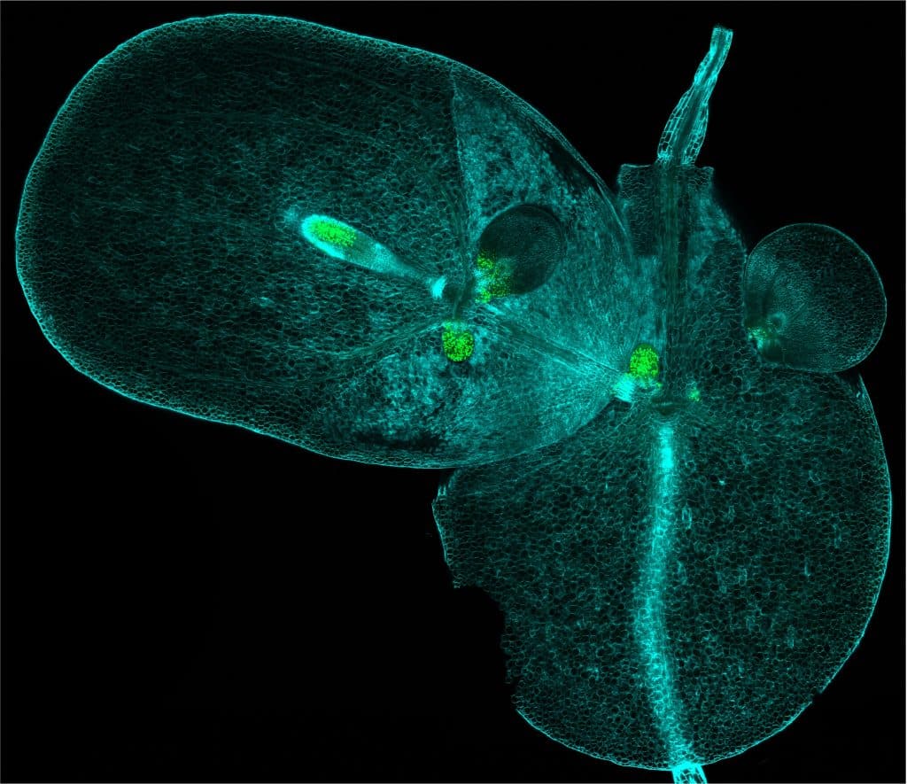
Our featured image, acquired by Emily Chan, shows a duckweed (Lemna minor) colony and the regions with actively dividing cells within the colony (coloured green).
In the colony shown here, there are a total of 21 individuals spanning five generations (mother, daughter, granddaughter, great-granddaughter, great-great-granddaughter). Although duckweeds are flowering plants, they primarily reproduce clonally. Progeny emerge from budding pouches of their mothers. The leaf-like structure is the frond body of a Lemna individual. Each Lemna individual bears a single root emerging from a region called the ‘node’, from which new daughters arise. In the literature, the duckweed ‘node’ is thought to be meristematic (i.e., a region containing self-renewing cells that supplies new cells for organogenesis, similar to animal stem cell niches). However, we know very little about what the ‘node’ is and what it does. This led us to investigate where the regions with active cell divisions in the duckweed body are found.
To achieve this, we labelled actively dividing cells in Lemna with 5-ethynyl-2’deoxyuridine (EdU), which intercalates DNA of actively dividing cells. We fixed the tissue with FAA (formaldehyde + acetic acid + ethanol), followed by a propanol wash to improve permeabilization, and then cleared the tissue with ClearSee, a clearing agent comprising urea, xylitol and sodium deoxycholate (Kurihara et al., 2015, Development; Nagaki et al., 2017, Scientific Reports). The Alexa Fluor 488 dye was conjugated to EdU molecules by click chemistry, enabling visualisation of cells with EdU labelling (coloured green in this image). The plant cell walls are stained with Calcofluor White, coloured blue in the image.
In this image, it is striking that only small, young duckweed fronds have active cell divisions, whereas the large, mature frond body no longer has active cell divisions.
Read more about Emily’s research.
Research career so far: I am fascinated by how developmental decisions occur on a cellular level for organ patterning and growth, and across various timescales in plants. Plants have a fundamentally different life strategy from animals. Plants do not move and their development is plastic since their body plan is determined in embryogenesis.
For my PhD, I studied petal patterning in the Hibiscus trionum flower in the Moyroud Group at the Sainsbury Laboratory, University of Cambridge. My PhD specifically was looking at the mechanisms of boundary formation using a combination of genetics, imaging and petal perturbations. As Hibiscus trionum is a relatively new model system, it was exciting to be able to do transgenics, which enabled us to test specific candidate genes. The Hibiscus trionum flower was beautiful to image on the microscope, as the bullseye pattern is composed of distinct colours, cell shapes and cell textures at the base and tip of the petal. I enjoyed tinkering with various forms of microscopy, including wide-field light microscopy, scanning electron microscopy, Raman microscopy and confocal microscopy.
Current research: Continuing a theme of studying developmental mechanisms in non-model plant species, I am now studying auxin signalling in duckweed in the Bishopp Group at the University of Nottingham. Duckweeds are aquatic plants and among the smallest and fastest growing plants on Earth. Duckweeds have an unusual body plan and have a highly simplified anatomy compared to most plants. We recently showed that duckweed shoot organs are compressed and fused, driving miniaturisation (Ware, Chan et al., 2026, Current Biology). A question we are trying to answer is, what drives the reduction in anatomical complexity in duckweeds? We found that duckweeds have a reduced number of signalling components of the plant hormone, auxin, which is a major regulator of plant growth and development. I am investigating whether a reduction in the number of components of the auxin signalling network in duckweed triggered a rewiring of gene regulatory networks controlling development.
Favourite imaging technique/microscope: My favourite imaging technique is probably confocal microscopy. It allows form to be linked to function. We can visualise the activity of various genes or proteins within the tissue using fluorescent proteins or dyes, which is very powerful. Confocal microscopes also have high resolution, and newer models are very good at multiplexing.
What are you most excited about in microscopy? I am most excited about adaptive imaging capabilities that are being developed. Microscopes with automated tracking capabilities enable us to follow growth of a tissue over longer periods as tissues often grow out of plane during the imaging time. Microscopes that can switch seamlessly between different imaging modalities on the same instrument are also a very exciting new development. This allows us to probe processes across different scales and correlate both fluorescent labels and label-free cellular context in the same sample without switching instrument.