Featured image with Joe McKellar
Posted by FocalPlane, on 8 December 2023
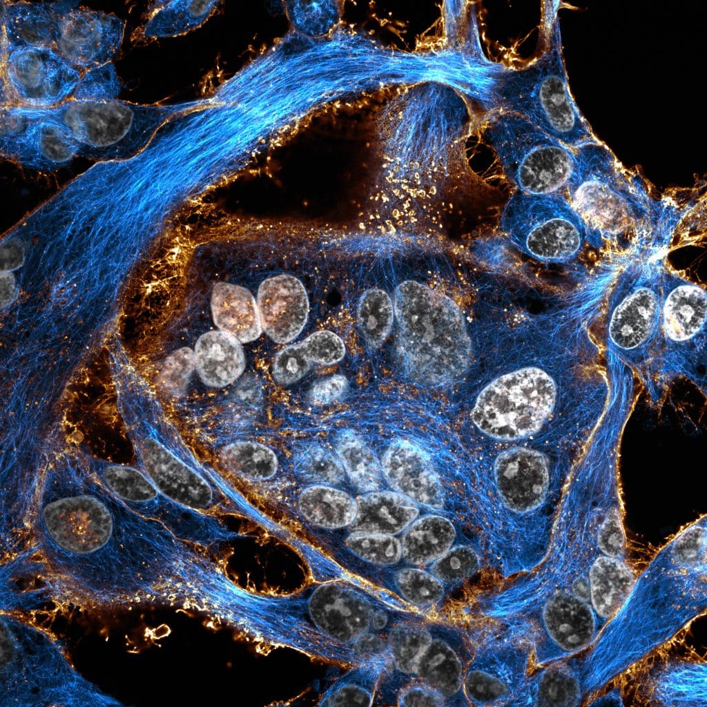
Our featured image shows a culture of boa constrictor kidney cells that have been transfected with a fusogenic viral protein. This leads to the formation of giant multi-nucleated cells. The cells were transfected with the fusogenic viral protein (shown in gold), left several days for efficient protein expression and for cell fusion to occur, plated onto coverslips, fixed and stained for nuclei (shown in gray) and microtubules (shown in cyan). The field of cells was acquired using the ZEISS confocal LSM980 microscope coupled to an Airyscan2 module. Deconvolution and post-processing were performed using the Zen Blue and FIJI software, respectively.

Find out more about Joe‘s research below:
Research career so far: My research career started with my Bachelor’s degree in Cell biology and Physiology at the University of Limoges in France. I then moved to the small city of Montpellier in the south of France, where I performed my Master’s degree in Microbiology and Immunology at the University of Montpellier. During this Master’s degree I started my wet lab work, with an internship in Dr Laurence Briant’s lab in the Institut de Recherche en Infectiologie de Montpellier (IRIM) where I worked on Chikungunya virus. My second Master’s internship was performed with Dr Caroline Goujon, also at the IRIM, and with whom I stayed to perform my PhD. During this second internship and PhD, I worked on trying to understand the mechanism of action of the antiviral MX1 protein against influenza A virus, with a focus on the cell biology of infection. It is during this time that I developed my love for microscopy. Following my PhD, I started a postdoc at the Institute for Molecular Genetics of Montpellier (IGMM) in Dr Karim Majzoub’s lab, where I am working on Deltaviruses.
Current research: Deltaviruses are the smallest known viruses to infect animals and have the particularity of being satellite viruses, meaning that they require the infection of another helper virus to form their own viral particles. Until recently, this family only contained the Hepatitis D virus (HDV), which associates with Hepatitis B virus (HBV) to produce its viral particles. However, recent studies have identified the existence of novel Deltaviruses that can replicate in rodents or even snakes! As of today, almost nothing is known about these viruses, and the helper viruses they require remain a mystery. Two recent studies have shown that Deltaviruses can associate with viruses of very different origins which therefore tempts the possibility of whether or not these viruses can host-shift, passing from animal to human populations. My research is currently focused on understanding the rules governing the association between Deltaviruses and their helper viruses and, in the long term, to determine the host-shifting potential of these enigmatic viruses.
Favourite imaging technique / microscope: My favourite imaging technique has to be classical confocal microscopy, although the confocal microscopes that I use allow for semi-superresolution possibilities, leading to higher resolution images! For these images, I use the Zeiss LSM880/980 microscopes that are coupled to Airyscan modules. I have spent countless hours in front of these two microscopes, and will spend many more!
What are you most excited about in microscopy? RNA viruses, for the most part, do not tolerate the insertion of large coding sequences in their genome, rendering the tagging of their viral proteins with fluorogenic proteins very difficult, bar the ectopic expression of tagged viral proteins in the infected cells themselves. I am very interested to see the further development of smaller and smaller fluorogenic tags, which may allow us to directly visualize the full viral lifecycle, from entry to egress!


 (No Ratings Yet)