Featured image with Marina Schernthanner
Posted by FocalPlane, on 16 August 2024
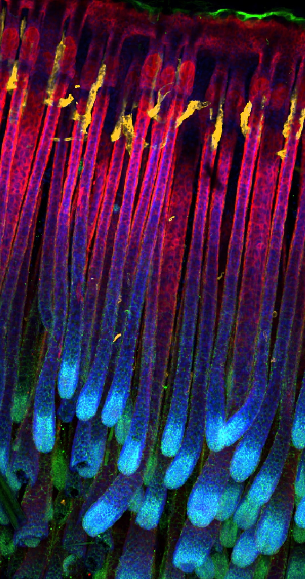
Our featured image, acquired by Marina Schernthanner, shows the hair follicles of a young (postnatal day 9) C57B6 mouse’s back skin. By using ethanol-based tissue clearing, Mariana stained fixed whole-mount tissue samples of the murine skin after removing the surface haircoat and subcutaneous fat layer. LYVE-1 (orange) marks the lymphatic vasculature, Keratin-14 (red) highlights the basal epidermal cells, P-Cadherin (green) outlines keratinocytes, and particularly the hair follicles, and DAPI (blue) marks the nuclei. Following clearing, the sample was imaged on an Andor Dragonfly spinning disc microscope (20x objective, tiled z-stacks). Image processing was done in Imaris 9.5.1.

Find out more about Marina’s research below:
Research career so far: I got my first glimpse into academic science when I was starting my master’s thesis work in Dr. Richard Flavell’s lab at Yale in 2017. It was also where I was introduced to the many fascinating aspects of working on the intestine. Back then, I was working on how prostaglandin signaling shapes intestinal health, branching into the areas of colorectal cancer and inflammatory bowel diseases. Most of my experimental skillset is rooted in what I learned in the Flavell lab, which equipped with me with a robust toolkit of techniques in molecular biology, immunology, tissue biology and cell culture. It also evoked a deep appreciation for basic science in me, with a specific knack for immunology. What followed were several 3-6 months-long research internships at the University of Innsbruck, where I did my undergrad, and the Cancer Institute of the Medical University in Vienna, exploring other areas (developmental biology) and tissues (bone marrow), before embarking on my PhD journey at the Rockefeller University in New York. Here, my enthusiasm for the intestine was re-ignited and I joined Dr. Elaine Fuchs’ laboratory to dig into the mechanisms of how stem cells in our barrier tissues – I am using the murine intestine and skin as my models – uphold tissue integrity and function.
Current research: For my PhD in Dr. Elaine Fuchs’ lab at the Rockefeller University, I am very interested in dissecting the modes of communication between tissue-resident stem cells (SCs) and the cells in their microenvironment (niche). In the lab we particularly focus on barrier tissues, which include the mammalian skin and intestine. Such barrier epithelia serve as an interface with the outside world, protecting organisms from pathogens and the like, while assimilating what is needed from the environment. In both the skin and the intestine, tissue-resident SCs fuel the efficient turnover of the epithelium or repair it upon an injury or insult. To do so, SCs heavily rely on input signals from neighboring cells . Using a paired transcriptomic approach we have recently profiled the interaction network between SCs and their niche in the mouse intestine. We exposed lymphatic capillaries as a new component of, and pivotal signaling hub in, the intestinal SC niche, where they orchestrate SC maintenance in the gut (1). Since then, I have continued to study how SCs engage in such crosstalk with their microenvironment, trying to draw parallels across barrier tissues and perhaps unveil conserved mechanisms of tissue homeostasis.
Favorite imaging technique/microscope: My favorite microscope in our lab is the Andor Dragonfly spinning disk – it is fast, has fantastic resolution and allows you to image a number of different samples. I have done multiplex imaging (IBEX) on mouse gut sections using this microscope and was always left in awe of the level of detail this microscope allowed me to capture.
What are you most excited about in microscopy currently/in the future? Speaking of IBEX, I’ve heard that the Germain lab, where this technique was originally developed, has a 3D version of multiplex imaging for whole-mount samples in the pipeline, so I am quite excited to learn how that works. Other than that, I am always mesmerized by in vivo live imaging studies to track cellular interactions in real-time and more recently, high-throughput approaches such as 10x Genomics’ Xenium platform to spatially map hundreds of cellular transcripts via imaging at single cell resolution.
References
1. Niec RE, Chu T, Schernthanner M, Gur-Cohen S, Hidalgo L, Pasolli HA, et al. Lymphatics act as a signaling hub to regulate intestinal stem cell activity. Cell Stem Cell. 2022;29(7):1067-82.e18.


 (No Ratings Yet)