Imaging with… Cellular Analysis Facility, University of Glasgow
Posted by FocalPlane, on 24 September 2025
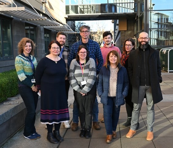
In our ‘Imaging with…’ blog post, we meet the staff of the Cellular Analysis Facility – Shared Research Facilities, College of Medicine, Veterinary & Life Sciences, University of Glasgow.
https://www.gla.ac.uk/colleges/mvls/shared-research-facilities/cellular-analysis
Staff role call
Leandro Lemgruber, Head of Cellular Analysis Facility
Expertise: light/electron microscopy, confocal/super-resolution, volumeEM, image analysis, tattoos and anything related to cats (yes, I want to see the photos of your cats!)
Most likely to be found running between the cores and different meetings, with my coffee flask!
Anthony Dornan, Deputy Head of Cellular Analysis Facility
Expertise: light-, confocal- and super-resolution- microscopy, image acquisition and analysis. A molecular geneticist that liked microscopes, now a microscopist that likes genetics.
Most likely to be foundcleaning up after users and writing passive/aggressive general notifications about oil usage on the systems.
Hannah Scales, Imaging Specialist/Lecturer
Expertise: In vivo/intravital imaging. Multiphoton microscopy; whole body imaging, imaging cytometry; high throughput microscopy; image acquisition and analysis. Teaching immunology, flow cytometry and microscopy. Immunologist and microscopist.
Most likely to be found trying to work out where I’m supposed to be and what I’m doing when I get there.
Susan Baillie, Senior Light Microscopy Support Technician
Expertise: light-, confocal- and super-resolution- microscopy,
Most likely to be found training/trouble shooting/chatting to our users. Persuading longer term users to leave the old microscopes behind and move to our new confocals!
Tyler Shaw, Histology Manager
Expertise: microtomy and cryotomy, immunohistochemistry and immunofluorescence, special staining, brightfield and fluorescence microscopy.
Most likely to be foundscraping paraffin off of everything, labelling slides and/or cassettes, and cleaning up spilt eosin
Margaret Mullin, Electron Microscopy Specialist/Lab Manager
Expertise: transmission and scanning electron microscopy, image acquisition, routine and specialised processing of biological samples, cryo-preparation techniques and Immunocytochemistry.
Most likely to be foundtraining, trouble shooting, chasing EM users’ precious samples, the size of microdots, around the bottom of an eppendorf tube, all the while wishing to be in my happy place on my EM microscopes exploring the wonders of biological world.
Diane Vaughn, Flow Cytometry Manager
Expertise: panel design, luminex assays, cell sorting, spectral and conventional analysis and data analysis.
Most likely to be found troubleshooting users experimental setup and/or data, or tinkering with a cytometer.
Alana Hamilton, Senior Flow Cytometry Support Technician
Expertise: conventional/spectral flow cytometry and cell sorting
Most likely to be found unblocking instruments, sending ‘Gentle Reminders’ and working on a spreadsheet of some form.
Daniel Lewis, Flow Cytometry Support Technician
Expertise: conventional flow cytometry and cell sorting
Most likely to be found training new users of flow cytometry analysis, and applying a spanner to an uncooperative cell sorter.
Microscope (and cytometer) role call:
Light Microscopy: Nikon AX-R NSPARC, Nikon A1-R, Zeiss Elyra7 Lattice SIM2, Zeiss LSM980 Multi-photon, Zeiss Upright LSM880, Zeiss Inverted LSM880, Zeiss Inverted LSM900, Zeiss CellObserver Spinning Disk, Zeiss PALM Laser Capture, Cytek ImageStream, ThermoScientific CX-5, ThermoScientific EVOS FL2 Auto, Leica DMi8.
Electron Microscopy: JEOL 1400Flash TEM, JEOL IT-100 SEM, Leica EM UC7 ultramicrotome (with cryochamber), Leica EM UCT ultramicrotome, Tousimis Autosamdri-815 crytical point dryer. Quorum Q150T metal coater, Leica EM GP2 plunge freezer, Gatan Elsa cryo-holder
Histology: Hamamatsu NanoZoomer-SQ Slide Scanner, Epredia Excelsior AS tissue processor, Histostart wax embedder, Epredia NX50 cryostat, Shandon Finesse 325 microtome.
Flow Cytometry: BD Canto II, BD Celesta, BD Fortessa, BD FACSAria II & III, Sony MA900, Sony ID7000, BD Symphony.
Who can access the facility?
While the Cellular Analysis Facility exists as part of the University of Glasgow’s College of Medicine, Veterinary and Life Sciences Shared Research Facilities (https://www.gla.ac.uk/colleges/mvls/shared-research-facilities/), access to the Facility’s equipment and expertise is available to all University staff, as well as associated companies, manufacturers and other academic institutes.
The Cellular Analysis Facility Imaging Suite is a purpose-built space, with specialised stations designed to accommodate fluorescence microscopy. Each station is networked and provisioned with electrical, compressed air and CO2 supply. The suite is designed to be able to accommodate new systems as they are acquired, as well as provide space for manufacturers to provide on-site demonstrations of new systems or for beta-testing developing technologies.
Facility staff are on hand to provide dedicated expert support and training in the office adjacent to the Imaging Suite. Additionally, the office has off-line workstations with Zeiss Zen and Nikon Elements licenses as well as specialist image analysis software platforms (Imaris, Huygens, Cell Profiler, Fiji) which are freely available for post-acquisition image processing and analysis by Facility users.
Flow Cytometry and Histology are located in dedicated units in the building adjoining the Imaging Suite. Electron Microscopy is again in a dedicated unit in a separate building on the University’s main campus.
The following section has been answered by Anthony Dornan (Deputy Head of Cellular Analysis Facility) and represents his personal opinion.
Pet peeve (something that users do that is annoying): Oil usage. Also, cleaning up the detritus of users once they’ve left the facility.
Favourite microscope: Currently, the Zeiss Elyra7 Lattice SIM2 (when it’s behaving) for its capacity for fine resolution of cellular processes and the Nikon AX-R NSPARC (when the Elyra isn’t behaving) for its multi-functional capabilities with respect to fast acquisition and resolution.
Favourite thing to image: Live cells – being able to visualise, and record, dynamic biological processes on a cellular, even molecular, scale remains astounding to me.
Best bit of advice (that you give or have been given): Best bit of advice I give to new users (I think) is, “the most expensive thing in this facility is your time – spend it wisely”.
Best bit of advice I was given when I came to the Facility, “Take some breaks and make sure you eat your lunch”.
If money was no object, we would buy… Light microscopy – Light sheet micro-system
EM – Volume EM system
Flow cytometry – Two new sorters – the Thinkcyte VisonSort, and either the BD or SONY Spectral sorter
Can you give us some examples of recent papers that were published with your assistance?
Synovial tissue myeloid dendritic cell subsets exhibit distinct tissue-niche localization and function in health and rheumatoid arthritis MacDonald L., Elmesmari A., Somma D., Frew J., Di Mario C., Madhu R., Paoletti A., Simakou T., Hardy O.M., Tolusso B., Campobasso D., Perniola S., Gessi M., Gigante M.R., Petricca L., Bruno D., Coletto L.A., Benvenuto R., Isaacs J.D., Filby A., McDonald D., Sim J.P.X., Jamieson N., Wei K., D’Agostino M.A., Millar N.L., Milling S., McSharry C., Gremese E., Affleck K., Baker K.F., McInnes I.B., Otto T.D., Korsunsky I., Alivernini S. and M. Kurowska-Stolarska. Immunity. 2024 Dec 10;57(12):2843-2862.e12. doi: 10.1016/j.immuni.2024.11.004.
Cancer-associated fibroblasts produce matrix-bound vesicles that influence endothelial cell function Santi A., Kay E.J., Neilson L.J., McGarry L., Lilla S., Mullin M., Paul N.R., Fercoq F., Koulouras G., Rodriguez Blanco G., Athineos D., Mason S., Hughes M., Thomson G., Kieffer Y., Nixon C., Blyth K., Mechta-Grigoriou F., Carlin L.M. and S. Zanivan. Sci Signal. 2024 Mar 12;17(827):eade0580. doi: 10.1126/scisignal.ade0580.
Two ancient membrane pores mediate mitochondrial-nucleus membrane contact sites Ovciarikova J., Shikha S., Lacombe A., Courjol F, McCrone R, Hussain W, Maclean A, Lemgruber L, Martins-Duarte ES, Gissot M and L. Sheiner. J Cell Biol. 2024 Apr 1;223(4):e202304075. doi: 10.1083/jcb.202304075.
Damage-associated cellular markers in the clinical and pathogenic profile of vaccine-induced immune thrombotic thrombocytopenia Abrams S.T., Du M., Shaw R.J., Johnson C., McGuinness D., Schofield J., Yong J., Turtle L., Nicolson P.L.R., Moxon C., Wang G., and C.H. Toh. J Thromb Haemost. 2024 Apr;22(4):1145-1153. doi: 10.1016/j.jtha.2023.12.008.
*Cover image for journal volume.
Compromised junctional integrity phenocopies age-dependent renal dysfunction in Drosophila Snakeskin mutants Dornan A.J., Halberg K.A., Beuter L.-K., Davies S.-A., and J.A.T. Dow. J Cell Sci 2023 136(19); jcs261118. doi:10.1242/jcs.261118.
*Cover image for journal volume also won the Nikon UK Microscopy Image Award 2023 & the Genetics Society of America 2024 Drosophila Image Award.
How should users acknowledge the facility and why is it important?
The idea of the Shared Research Facilities is to ensure that, logistically, systems are being used as efficaciously as possible, while researchers, both staff and students, are being provided with expert support and guidance to ensure that they achieve their research goals. Scientific publications provide a key measure for how successful the Facility is in achieving this aim of advancing research projects, both within and outside the University of Glasgow. As such, students and staff are encouraged to credit the input that the Facility has had on their research, from a simple acknowledgment to, where appropriate, co-authorships. This need is also communicated to students, staff, PI’s and College management through regular showcases of the Facility’s resources and capabilities, using real examples of research and imaging generated by Facility users and staff.
 (No Ratings Yet)
Get involved
Create an account or log in to post your story on FocalPlane.
More posts like this

Filter by
- NewsApply
- DiscussionsApply
- How toApply
- ToolsApply
- Case studiesApply
- InterviewsApply
- JobsApply
- EducationApply
- Blog seriesApply
- Deep Learning for Bi..o-image analysisApply
- GloBIAS – updates fr..om the communityApply
- WAMBIAN: West Africa.. in FocusApply
- Volume EMApply
- Latin American Micro..scopistsApply
- Bio-image Analysis w..ith NapariApply
- Imaging with…Apply
- Towards Global Acces..sApply
- Latin America Bioima..gingApply
- From Zero to Qupath ..HeroApply
- Asian Microscopists ..and Cell BiologistsApply
- AIC at HHMI JaneliaApply
- Highlights from Euro..-BioImagingApply
- LSFM seriesApply
- DIY MicroscopyApply
- View all