Microscopy preprints – new tools and techniques in imaging
Posted by FocalPlane, on 5 September 2025
Here is a curated selection of preprints posted recently on new tools and techniques in imaging. Let us know if we are missing any recent preprints that are on your reading list!
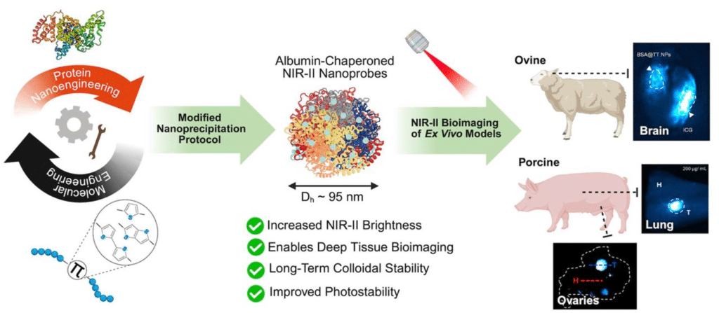
Ultrabright NIR-II Nanoprobes for Ex Vivo Bioimaging: Protein Nanoengineering Meets Molecular Engineering
Isabella Vasquez, Asma Harun, Robert Posey, Ruhan Reddy, Ulrich Bickel, Joshua Tropp, Indrajit Srivastava
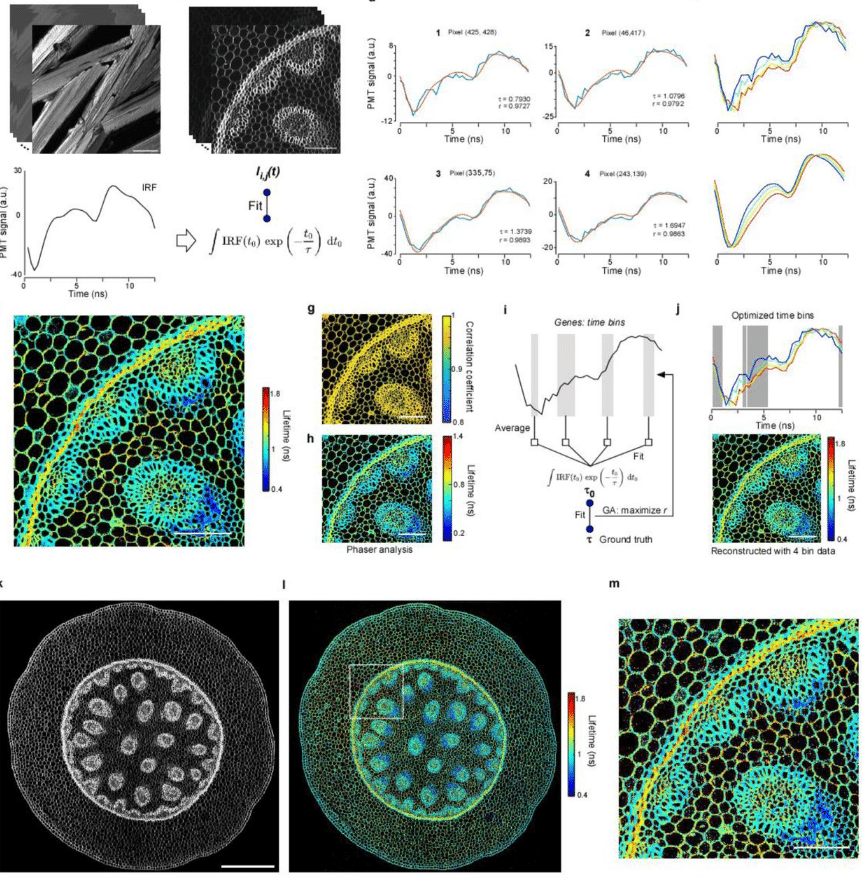
Single-dye, transfection-free FLIM multiplexing via bioorthogonal chemistry
Neville Dadina, Justin H. Kwon, Lauren Lesiak, Shuai Zheng, Madeline Zoltek, Daniel Brauer, Alanna Schepartz
Using DNA origami to study nanoscale organization of plasma membranes
Eloina Corradi, Konlin Shen, Zeynep Karatas, Maureen Cercy, Thomas Schlichthaerle, Melissande Osouf, Brune Vialet, Philippe Barthelemy, Morgane Rosendale, Adiyodi Veetil Radhakrishnan, Tianchi Chen, Ralf Jungmann, Arnaud Gissot, Shawn Douglas, Grégory Giannone
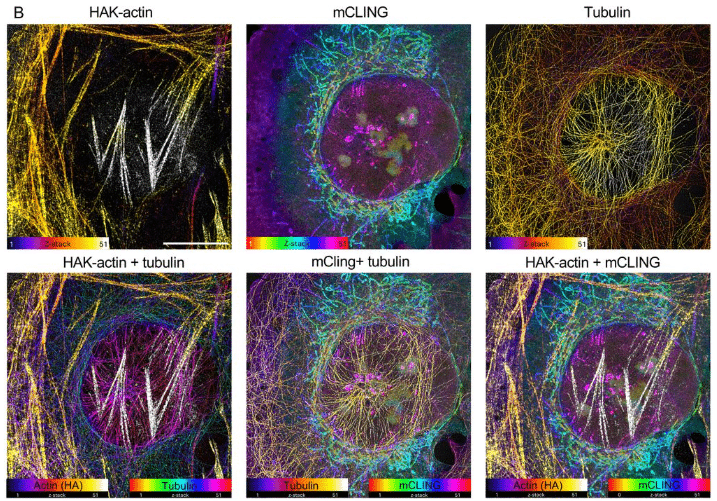
HAK-actin, U-ExM-compatible probe to image the actin cytoskeleton
Olivier Mercey, Luc Reymond, Florent Lemaître, Isabelle Mean, Marine H. Laporte, Marine Olivetta, Karin Sadoul, Omaya Dudin, Virginie Hamel, Paul Guichard
Super-resolution live-cell mapping of protein-protein interactions using chemogenetic split reporters and STED microscopy
Stephanie Board, Arnaud Gautier
Absolute Membrane Potential Recording with ASAP-Type Genetically Encoded Voltage Indicators Using Fluorescence Lifetime Imaging
Anagha Gopalakrishnan Nair, Marko Rodewald, Hyeonsoo Bae, Philipp Rühl, Jürgen Popp, Michael Schmitt, Tobias Meyer-Zedler, Stefan H. Heinemann
Three-dimensional multi-target super-resolution microscopy of cells using Metal-Induced Energy Transfer and DNA-PAINT
Nazar Oleksiievets, Nikolaos Mougios, Samrat Basak, Daniel C. Jans, Lara Hauke, Jan Christoph Thiele, Stefan Jakobs, Felipe Opazo, Jörg Enderlein, Roman Tsukanov
Automated and Simulation-Guided Multiplexed DNA-PAINT for Nanoparticle Characterization
Stijn van Veen, Emiel W. A. Visser, Lorenzo Albertazzi
Simulation-Guided Exploration of PAINT Parameter Space for Accurate Molecular Quantification
Wei Shan Tan, Arthur M. de Jong, Menno W. J. Prins
Single-Cell Super-Resolution Quantification of Oxidative DNA Damage via Aptamer-Assisted DNA-PAINT
Jingfang Zhao, Li-Sheng Zhang, Yibin Liu, Shuoxing Jiang, Limin Xiang
Assessing cellular metabolic dynamics with NAD(P)H fluorescence polarization imaging
Lu Ling, Jack C. Crowley, Matthew L. Tan, Jennie A.M.R Kunitake, Adrian A. Shimpi, Rebecca M. Williams, Lara A. Estroff, Claudia Fischbach, Warren R. Zipfel
A Suite of Stains: Characterization of four fluorophores as complementary tools for visualizing neutral lipids in an extremophilic green alga
Pomona Osmers, Eliza-Jayne Y. Boisvert, Christopher N. Boddy, Deryn E. Fogg, Marina Cvetkovska
Real-time feedback control microscopy for automation of optogenetic targeting
Lucien Hinderling, Alex E. Landolt, Benjamin Grädel, Laurent Dubied, Cédric Zahni, Moritz Kwasny, Agne Frismantiene, Talley Lambert, Maciej Dobrzyński, Olivier Pertz
Smart Microscopy: Current Implementations and a Roadmap for Interoperability
Lucien Hinderling, Hannah S. Heil, Alfredo Rates, Philipp Seidel, Manuel Gunkel, Benedict Diederich, Thomas Guilbert, Rémy Torro, Otmane Bouchareb, Claire Demeautis, Célia Martin, Scott Brooks, Evangelos Sisamakis, Grandgirard Erwan, Karl Johansson, Johannes K. Ahnlinde, Oscar André, Philip Nordenfelt, Pontus Nordenfelt, Claudia Pfander, Jürgen Reymann, Talley Lambert, Marco R. Cosenza, Jan O. Korbel, Rainer Pepperkok, Lukas C. Kapitein, Olivier Pertz, Nils Norlin, Aliaksandr Halavatyi, Rafael Camacho
Pan-ASLM: a high-resolution and large field-of-view light sheet microscope for Expansion Microscopy
Hannahmariam T. Mekbib, Lasse Pærgård Andersen, Shuwen Zhang, Jonathan Gulcicek, Yuan Tian, Jack R. Ross, Mark D. Lessard, Joerg Bewersdorf
Deep-learning-assisted SICM for enhanced real-time imaging of nanoscale biological dynamics
Z. Ayar, M. Penedo, B. Drake, J. Shi, S. M. Leitao, I. Krawczuk, H. Miljkovic, A. Radenovic, J. Ban, V. Cevher, G. E. Fantner
Superresolution imaging of live samples by centroid reassignment microscopy
Chuan Li, Quan Le, Julian O. Kimura, Bingying Zhao, Yunzhe Li, Jian Zhao, Thomas Bifano, Brandon Weissbourd, John T. Ngo, Jerome Mertz
Combining Multi-site FRAP and HILO-TIRF microscopy using a Spatial Light Modulator
Avinash Upadhya, Yean Jin Lim, Woei Ming Lee
Event-triggered MINFLUX microscopy: smart microscopy to catch and follow rare events
Jonatan Alvelid, Agnes Koerfer, Christian Eggeling
Real-time, High-throughput Super-resolution Microscopy via Panoramic Integration
Kyungduck Yoon, Hansol Yoon, Kidan Tadesse, Zhi Ling, Biagio Mandracchia, Sayantan Datta, G Ozan Bozdag, Anthony J Burnetti, William C Ratcliff, Shu Jia
Learned Single-Pixel Fluorescence Microscopy
Serban C. Tudosie, Valerio Gandolfi, Shivaprasad Varakkoth, Andrea Farina, Cosimo D’Andrea, Simon Arridge
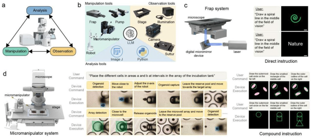
Embodied Intelligence Unlocks Autonomous Microscopy
Gang Huang, Zhengyang Zhang, Songlin Zhuang, Yang Wu, Zhihui Lu, Mingsi Tong, Huijun Gao
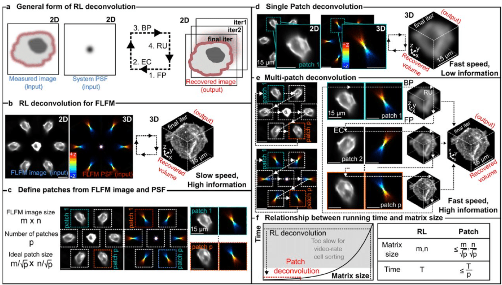
Patch deconvolution for Fourier light-field microscopy
Bin Fu, Caroline L. Jones, Daniel Heraghty, Shengbo Yang, Caitlin O’Brien-Ball, Victoria Junghans, Haowei Yang, Tuomas P.J. Knowles, Lucien E. Weiss, Ricardo A. Fernandes, Steven F. Lee
Single Capture Quantitative Oblique Back-Illumination Microscopy
Paloma Casteleiro Costa, Srinidhi Bharadwaj, Zhenmin Li, Nischita Kaza, Mercedes Lopez-Esteva, Anthony Lien, Bilal Haider, Francisco E. Robles
An open, integrated platform for multiplexed bioluminescence microscopy
Lorenzo Scipioni, Belen Torrado, Giulia Tedeschi, Lila P. Halbers, Zachary R. Torrey, Erin B. Fuller, Francesco Fersini, Christoph Gohlke, Andrej Luptak, Jennifer A. Prescher, Michelle A. Digman
Spinning disk confocal microscopy with a 25 Megapixel Camera
Guy M. Hagen, Brian Lewis, Summer Levis, Joseph R. Hamilton, Tristan C. Paul
Refractive index mapping below the diffraction limit via single molecule localization microscopy
Simon Jaritz, Lukas Velas, Anna Gaugutz, Manuel Rufin, Philipp J. Thurner, Orestis G. Andriotis, Julian G. Maloberti, Simon Moser, Alexander Jesacher, Gerhard J. Schütz
Super-resolved live imaging of thick biological samples with 3D Random Illumination Microscopy (3D-RIM)
Thomas Mangeat, Lorry Mazzella, Benoît Rogez, Guillaume Giroussens, Mathilde Bernard, Pablo Vargas, Marc Allain, Simon Labouesse, Jérôme Idier, Loïc LeGoff, Anne Sentenac
Separate-scan atomic force microscope for fast infrared scattering-type scanning near-field optical microscope
Yusuke Sakiyama, Santiago H. Andany, Georg E. Fantner, Joachim Heberle
4-Pi Stimulated Raman Scattering for Label-free Super-resolution Chemical Imaging
Jonathan I. Kim, Zachary Ellsworth, Erin L. Dunnington, Nidhi R. Mehta, Chisa Zensho, Dan Fu
Voltage Imaging with Periodic Structured Illumination
Forest Speed, Alec Teel, Gregory L. Futia, Diego Restrepo, Emily A. Gibson
Pulsed-electron illumination does not reduce beam damage for imaging biological macromolecules
Vishal Kumar, Julika Radecke, K.V. Chinmaya, Inayathulla Mohammed, Ricardo C. Guerrero-Ferreira, Daniel Harder, Dimitrios Fotiadis, Henning Stahlberg
Systematic Characterization of Optical Aberrations Reveals Cryo-FLM Localization Fidelity
Hongjia Li, Lauren Ann Metskas, Fang Huang
High speed functional imaging with a microfluidics-compatible open-top light-sheet microscope enabled by model predictive control of a tunable lens
W. Alexander Calhoun, Sihoon Moon, Lucinda Peng, Hang Lu
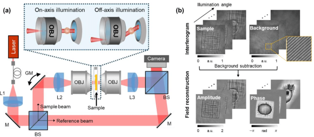
Inverse-scattering in biological samples via beam-propagation
Jeongsoo Kim, Blythe Bolton, Khashayar Moshksayan, Rishika Khanna, Mary E. Swartz, Michał Ziemczonok, Mohini Kamra, Karin A. Jorn, Sapun H. Parekh, Małgorzata Kujawińska, Johann Eberhart, Elif Sarinay Cenik, Adela Ben-Yakar, Shwetadwip Chowdhury
Single-molecule flow cytometry
Amir Rahmani, Matthew Christie, Amy Truesdale, James Thorne, Aleks Ponjavic
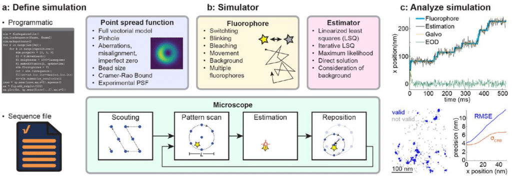
Evaluating MINFLUX experimental performance in silico
Zach Marin, Jonas Ries
Low Angle Ring Illumination Stereomicroscopy (LARIS): An improved imaging method of Drosophila compound eyes
Jukta Biswas, Ankur Kumar, Anand K Singh
A hierarchical adaptive optics strategy for three-photon imaging during behavior
Huriye Atilgan, Jingyu Wang, Qi Hu, Sandra Tan, Blake Russell, Randy M. Bruno, Martin J. Booth, Armin Lak
Open-source modular FPGA system for two-photon mesoscope enabling multi-layer, multi-depth neural activity recording and lifetime imaging
Riichiro Hira, Fumiya Imamura, Hiroto Imamura, Yuki Yoneyama, Takehisa Handa, Osamu Fujioka, Che-Hang Yu, Satoshi Suitoh, Reiko Hira, Atsushi Kamoshida, Shigeki Kato, Kazuto Kobayashi, Hiroki Shiwaku, Hidehiko Takahashi, Spencer L. Smith, Akihiro Funamizu, Yoshikazu Isomura
Streamlined Montage Cryo-Electron Tomography for Exploring the Ultrastructure of Cells and Tissues
Ryan Hylton, Micaela Boiero Sanders, Adriana Prajica, Gavin Rice, Stefan Raunser
Miniaturized widefield microscope for high speed in vivo voltage imaging
Catherine A. Saladrigas, Forest Speed, Alec Teel, Mo Zohrabi, Eduardo J. Miscles, Gregory L. Futia, Larry V. Baker, Ye Zhang, Ioannis Kymissis, Victor M. Bright, Cristin G. Welle, Diego Restrepo, Juliet T. Gopinath, Emily A. Gibson
High-throughput 3D super-resolution ultrasound imaging
Weisong Zhao, Nanchao Wang, Zhenqian Han, Xining Xu, Xiangyu Ma, Tianhua Zhou, Jiahui Gui, Yuzhen Liu, Qianbo Liu, Liying Qu, Wenhao Liu, Xiangyan Ding, Xin Liu, Dean Ta, Jiubin Tan, Liangyi Chen, Junjie Yao, Haoyu Li
Optimal tilt-increment for cryo-ET
Maarten W. Tuijtel, Tomáš Majtner, Beata Turoňová, Martin Beck
Cryo-EM protein structure without purification
Samantha M. Powell, James E. Evans
VitriFlex: An Open-Source, Modular, and Customizable Robotic Platform for Cryo-EM Grid Preparation
Wyatt Peele, Kedar Sharma, Thomas B. Stanley, Randy K. Bledsoe, Robert M. Petrovich, Mario J. Borgnia, Venkata P. Dandey
 (No Ratings Yet)
Get involved
Create an account or log in to post your story on FocalPlane.
More posts like this

Filter by
- NewsApply
- DiscussionsApply
- How toApply
- ToolsApply
- Case studiesApply
- InterviewsApply
- JobsApply
- EducationApply
- Blog seriesApply
- Asian Microscopists ..and Cell BiologistsApply
- AIC at HHMI JaneliaApply
- Deep Learning for Bi..o-image analysisApply
- GloBIAS – updates fr..om the communityApply
- WAMBIAN: West Africa.. in FocusApply
- Volume EMApply
- Latin American Micro..scopistsApply
- Bio-image Analysis w..ith NapariApply
- Imaging with…Apply
- Towards Global Acces..sApply
- Latin America Bioima..gingApply
- From Zero to Qupath ..HeroApply
- Highlights from Euro..-BioImagingApply
- LSFM seriesApply
- DIY MicroscopyApply
- View all