Imaging spotlight: Initiation of chromosome congression deconstructed by multi-scale live lattice light-sheet and super-resolution microscopy
Posted by Kruno Vukušić, on 16 January 2026
In this highlight, Kruno Vukušić and Iva M. Tolić describe how they use multi-scale live lattice light-sheet and super-resolution microscopy to decipher the earliest steps of chromosome congression. Chromosome congression is a central early step in mitosis that ensures accurate chromosome segregation. In two recent studies published in Nature Communications (here and here), they set out to understand how initiation operates at both mechanical and molecular levels, and particularly how communication between kinetochores and centrosomes regulates these earliest steps. Addressing this question required imaging approaches capable of capturing fast and transient events within the dense and highly dynamic environment of the mitotic spindle.
- What was the main focus of this work and what did you find?
Our work focused on understanding how chromosome congression is initiated, a fast and dynamic process essential for accurate segregation. Previously, initiation was not the main focus because kinetochore motor CENP-E was primarily thought to act as a transport motor moving polar chromosomes toward the spindle equator. By integrating multiple imaging and experimental approaches, we introduced the concept of movement initiation and found that CENP-E plays a key role by promoting rapid stabilization of initial end-on kinetochore–microtubule attachments through antagonism of Aurora kinase activity. Further control of timing and location of initiation is provided by kinetochore–centrosome feedback. Once initiation has occurred, subsequent chromosome movements proceed with similar dynamics even if CENP-E activity is reduced.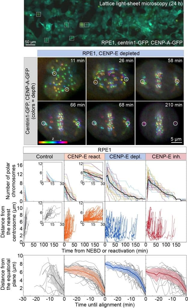
Figure 1. Lattice light-sheet microscopy of chromosome alignment dynamics following CENP-E perturbation. Lattice light-sheet microscopy (LLSM) field of view showing RPE-1 cells expressing centrin1-GFP and CENP-A-GFP, colour-coded by depth. Cells were treated with 80 nM CENP-E inhibitor (GSK-923295) for 3 h prior to imaging; boxed regions indicate mitotic cells (top). Representative time-lapse images show an RPE-1 cell after CENP-E depletion (middle). All images are maximum-intensity projections; time 0 marks mitotic entry. Quantification shows the number of polar chromosomes (bottom, top), their distance to the nearest spindle pole (bottom, middle), and their distance from the equatorial plane until successful alignment (bottom, bottom) for the indicated treatments. Thick lines indicate means; shaded regions show standard deviation. - Which imaging techniques were essential for this work and why?
To capture the earliest steps of congression, we required imaging that combined speed, scale, and low phototoxicity. Lattice light-sheet microscopy was our primary method, enabling fast three-dimensional live-cell imaging with large fields of view and whole-cell coverage. This allowed long-term observation of many mitotic cells in parallel and the capture of many rare, short-lived initiation events. Confocal microscopy was used for dynamic live assays to test multiple inhibitors, providing higher throughput. The mitotic spindle is a highly dense structure composed of many closely packed microtubules, which limits the resolution achievable with diffraction-limited microscopy. To visualize spindle architecture and kinetochore-associated structures in detail, we complemented live imaging with super-resolution microscopy. In particular, STED microscopy was essential to resolve microtubule organization at kinetochores, fibrous corona dynamics, and recruitment of end-on attachment markers. This multimethod approach allowed us to integrate information across multiple scales of spindle organization and chromosome movement dynamics. - How did you capture such fast and transient events experimentally, and how was analysis performed?
A key strategy was acute chemical inhibition of CENP-E followed by washout to reactivate the motor. This slowed and temporally constrained initiation of congression, compressing initiation events into a defined time window. This allowed us to observe initiation at time-scales compatible with long lattice light-sheet experiments and to capture many events in a single imaging session. Similar experimental tricks could in principle be applied to other molecular motors, not just mitotic events. For analysis, we used semi-automated image analysis pipelines to process large three-dimensional time series and track centrosomes and kinetochores. Automated detection was combined with manual curation, necessary because kinetochore movements can be sudden and trajectories often cross in the dense spindle environment. These analyses enabled quantitative measurements of chromosome position, movement trajectories, timing of congression initiation, and recruitment of end-on attachment markers. - What were the main technical challenges and lessons learned?
Lattice light-sheet microscopy routinely generates terabyte-scale datasets per experiment, making data handling a major challenge. Successful projects require early planning of storage, data transfer, and processing pipelines, as well as collaboration with institutional and national high-performance computing infrastructure. Semi-automated tracking was effective but labor-intensive, and fully automated tracking of dense kinetochore populations remains difficult. Developing robust end-to-end tracking algorithms for large 3D time-lapse datasets is an important next step for the field.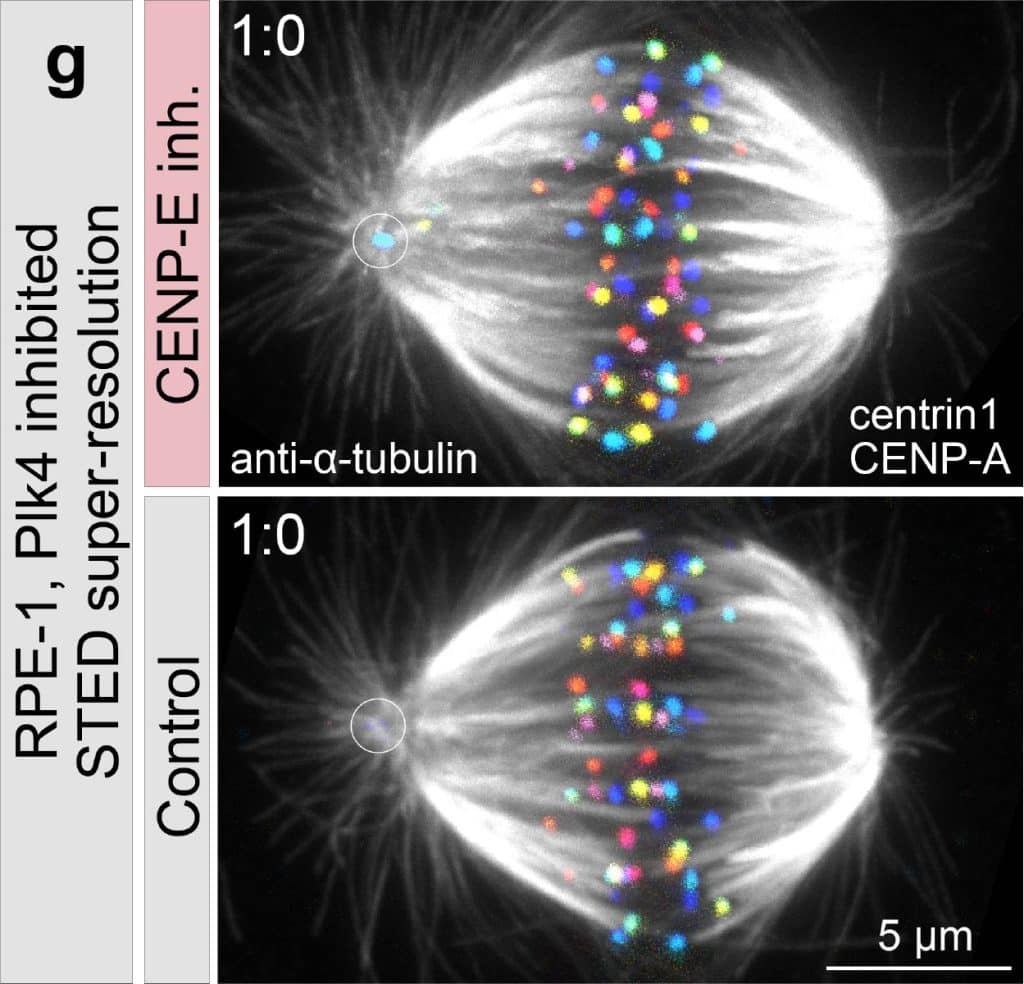
Figure 2. STED microscopy of mitotic RPE-1 cells. RPE-1 cells treated with 300 nM centrinone were imaged with (top) or without (bottom) 80 nM CENP-E inhibitor (GSK-923295). Cells were immunostained for α-tubulin (grey) and express CENP-A–GFP and centrin1–GFP, color-coded by depth. All images show maximum-intensity projections. - Where can people find more information?
Primary research articles:- Vukušić K, Tolić IM. CENP-E initiates chromosome congression by opposing Aurora kinases to promote end-on attachments. Nature Communications. 2025;16:8537. doi:10.1038/s41467-025-64148-w.
- Vukušić K, Tolić IM. Kinetochore–centrosome feedback linking CENP-E and Aurora kinases controls chromosome congression. Nature Communications. 2025;16:9097. doi:10.1038/s41467-025-64804-1.
Key imaging and methods resources from our lab:- Koprivec I, Štimac V, Tolić IM. Super-resolution imaging of mitotic spindle microtubules using STED microscopy. In: The Mitotic Spindle. Methods in Molecular Biology, vol. 2872. Springer; 2025:3–19. doi:10.1007/978-1-0716-4224-5_1.
- Ponjavić I, Vukušić K, Tolić IM. Expansion microscopy of the mitotic spindle. Methods in Cell Biology. 2021;161:247–274. doi:10.1016/bs.mcb.2020.04.014.
 (No Ratings Yet)
Get involved
Create an account or log in to post your story on FocalPlane.
More posts like this

Filter by
- NewsApply
- DiscussionsApply
- How toApply
- ToolsApply
- Case studiesApply
- InterviewsApply
- JobsApply
- EducationApply
- Blog seriesApply
- Volume EMApply
- Latin American Micro..scopistsApply
- Bio-image Analysis w..ith NapariApply
- Imaging with…Apply
- Towards Global Acces..sApply
- Latin America Bioima..gingApply
- From Zero to Qupath ..HeroApply
- Asian Microscopists ..and Cell BiologistsApply
- AIC at HHMI JaneliaApply
- Deep Learning for Bi..o-image analysisApply
- GloBIAS – updates fr..om the communityApply
- Highlights from Euro..-BioImagingApply
- LSFM seriesApply
- DIY MicroscopyApply
- View all