

Pearling and directed trafficking as determinants of mitochondrial genome organization and network architecture
Posted by FocalPlane, on 21 July 2026
In this highlight, we hear from Juan C. Landoni, who discusses his recent work uncovering how mitochondria distribute their genome through pearling (Landoni et al. 2026, Science), and the key role of mitochondrial trafficking in maintaining new material dissemination and network architecture (Winter, Landoni et al. 2026, JCB).
What are the key results from your papers?
Both papers uncover spatial organization principles in mitochondrial dynamics that had remained largely undescribed, owing to both technological limitations in imaging, as well the field’s predominant focus on fission-fusion dynamics.
The Science paper addresses a long-standing fundamental question in mitochondrial biology: how is the mitochondrial genome spatially regulated? Mitochondrial DNA nucleoids are maintained at remarkably regular spacing, which is fundamental for their inheritance and function. We discovered that the mechanism enabling this is pearling, a spontaneous and reversible transition from tubular to beads-on-a-string morphology driven by a Rayleigh-Plateau-like biophysical instability. The characteristic length scale imposed by pearling matches inter-nucleoid spacing, and the transition itself simultaneously disaggregates multi-copy nucleoids and redistributes them at regular intervals. We found that pearling is triggered by calcium influx at ER-mitochondria contact sites. Lamellar cristae, in turn, provide the elastic resistance that controls its probability and, after recovery, sterically confine nucleoids to prevent re-merging.
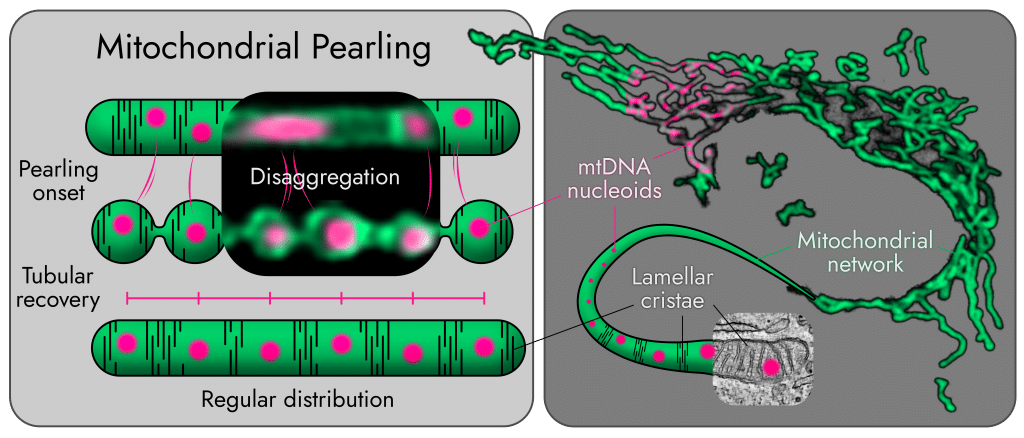
The JCB paper, led by Julius Winter, investigates the interplay between the spatial organization of mitochondrial biogenesis, trafficking, and network architecture. Mitochondrial trafficking has been historically investigated in highly polarized cells like neurons, but mostly overlooked in morphologically simpler cell types. Combining quantitative imaging with cellular micropatterning and in silico modelling (in collaboration with Elena Koslover and Keaton Holt from UCSD), we found that mitochondrial transport is strongly anterograde-biased from the perinuclear area, distributing newly synthesized material from the perinuclear biogenesis hub to the periphery. Intriguingly, this asymmetric flux is required to maintain both network mass distribution and connectivity across the cell.
What imaging techniques have you used in your research?
The projects required a truly large array of imaging approaches, not simply because imaging is fun and exciting (which we can all agree it is), but because the biological questions demanded it.
Pearling events happen right below the diffraction limit, meaning we needed extended- or super-resolution to capture them accurately, while simultaneously requiring fast imaging as they are reversible within seconds. We also needed to investigate the detailed ultrastructure of cristae during pearling events (well below the diffraction limit) at comparable speeds, while quantifying their frequency and impact across entire cells over longer periods, and whenever possible obtaining quantitative fluorescent signal without non-linear computational processing. All this while minimizing phototoxic stress on the cells. No single modality could provide all the evidence we needed. In response, we adapted the microscopy to each biological question and, when possible, confirmed observations with multiple imaging paradigms spanning varying spatial and temporal resolution and phototoxicity tradeoffs.
Similar challenges arose in the trafficking work, where mitochondrial material needed to be accurately tracked over more than an hour and associated with fast, seconds-long runs as well as longer processes such as mtDNA replication and protein translation. Combining photoconversion of mitochondria with cellular micropatterning provided a quantitative platform to compare across many cells with typically different morphologies, and to obtain robust data for otherwise elusive processes.
Are there technical tips or tricks that you have learnt while doing this research?
Phototoxicity: Whenever possible we used short-term imaging and the lowest irradiation compatible with the needed resolution, as described above. We also explored label-free phase contrast imaging and “smart” adaptive acquisition to capture events only when detected by machine learning.
Choice of probes: Nucleoid markers are notorious for disrupting mtDNA homeostasis, which made investigating subtle changes particularly tricky. Each probe carries its own artefact profile: DNA-intercalating dyes are potentially mutagenic over time, and the packaging protein TFAM fused to fluorophores is documented to affect nucleoid packaging and gene expression. Using several probes, each imperfect, allowed us to draw stronger conclusions from their collective outputs.
Modelling: In silico modelling did not merely provide a way to simulate the data. By informing the models with experimental values, we observed unexpected outcomes that in turn allowed us to reassess previously confusing data and redesign experiments to corroborate. The bidirectional conversation between modelling and experiment was invaluable.
What were the key image analysis lessons from these projects, and what should others working on mitochondrial dynamics watch out for?
Quantitative biology requires both creativity and rigor. AI-driven approaches can be powerful, but if we train a model on what we expect it to find, we remain blind to the unexpected.
Both projects required developing innovative ways of quantifying phenomena, as no existing approaches were adequate. Often this involved extensive manual labeling and blinded classification to obtain quality data while minimizing bias, and carefully evaluating (and resisting) the urge to invest large amounts of time in using or developing automated pipelines. While automation is very powerful, one should not underestimate how time-consuming it can be compared to robust “traditional” quantification, and existing tools are typically optimized for well known processes. The later constrains one’s analysis existing paradigms, missing what lies outside their design assumptions. There is no one right approach, but innovatively combining many while taking the time to think critically about their trade-offs always pays off.
Quantifying material spreading from mitochondria was one example with creative approaches for quantification. After many attempts, displaying the signal as a normalized kymograph of the integrated central axis of the cell revealed a clear process which was invisible to other characterization attempts, and robustly quantifiable. Constructive reviewer feedback then allowed us to expand the findings further, using experimental thresholds of mitochondrial morphology and spreading to perform radial quantification analyses on independent datasets, unlocking our ability to also quantify non-patterned cells. Pearling frequency was another key example: a critical measure with non-trivial quantification issues. Events per minute was intuitive but ignored both the magnitude of individual events and the total mitochondrial content in the cell, while more thorough normalization approaches required increasingly challenging and error-prone segmentation pipelines. After extensive reflection and testing, we settled on the percentage of total network length that pearls at least once per minute, measuring both pearling extent and network as skeletonized length. No approach will ever be perfect, but this was the best combination of robustness, minimized error and bias, consistent normalization, and biological interpretability.
Are there any advances in imaging or image analysis that would help your research going forward?
I believe the imaging field is moving in a great direction for biological discovery, particularly in developing imaging systems that extend resolution while still enabling physiological imaging with minimal phototoxicity. On the analysis side, more and more pipelines are emerging to segment and quantify large datasets, but the landscape remains either fully automated or fully manual. Approaches that keep a human in the loop, accessible to biologists with limited programming expertise while still enabling hybrid data analysis, would substantially accelerate the field.
More broadly, thinking across disciplines and outside the box matters. Technological development in microscopy is proceeding at an unprecedented pace, but communication between technology developers and biologists driving new discovery remains limited. As a consequence, novel platforms are repeatedly validated on familiar structures as proof of principle, while remaining inaccessible to end users because they seem intimidating. Interdisciplinary collaboration within diverse teams goes a long way.
Get involved
Create an account or log in to post your story on FocalPlane.
More posts like this

Filter by
- NewsApply
- DiscussionsApply
- How toApply
- ToolsApply
- Case studiesApply
- InterviewsApply
- JobsApply
- EducationApply
- Blog seriesApply
- Volume EMApply
- Latin American Micro..scopistsApply
- Bio-image Analysis w..ith NapariApply
- Imaging with…Apply
- Towards Global Acces..sApply
- Latin America Bioima..gingApply
- From Zero to Qupath ..HeroApply
- Asian Microscopists ..and Cell BiologistsApply
- AIC at HHMI JaneliaApply
- Deep Learning for Bi..o-image analysisApply
- GloBIAS – updates fr..om the communityApply
- WAMBIAN: West Africa.. in FocusApply
- Highlights from Euro..-BioImagingApply
- LSFM seriesApply
- DIY MicroscopyApply
- View all