

Imaging spotlight: Live imaging reveals epithelial cell extrusion in a sea anemone
Posted by Ines Fournon-Berodia, on 7 September 2026
In this Imaging spotlight, Inés Fournon Berodia and colleagues share how live confocal imaging and machine-learning-assisted image analysis revealed epithelial cell extrusion in the sea anemone Nematostella vectensis.
Nematostella can dramatically shrink its body during starvation by losing large numbers of cells. We wanted to understand how this was possible in shrinking polyps without compromising the barrier function of their epithelia. The key challenge was to see what was happening to individual epithelial cells in the epidermis.
What are the key results from your paper?
A major part of this work was developing a way to image the epidermis of whole, live juvenile Nematostella polyps at subcellular resolution. Previous live imaging in Nematostella had largely focused on embryos, which are considerably smaller and easier to immobilise. Imaging a juvenile animal presented a very different technical challenge.
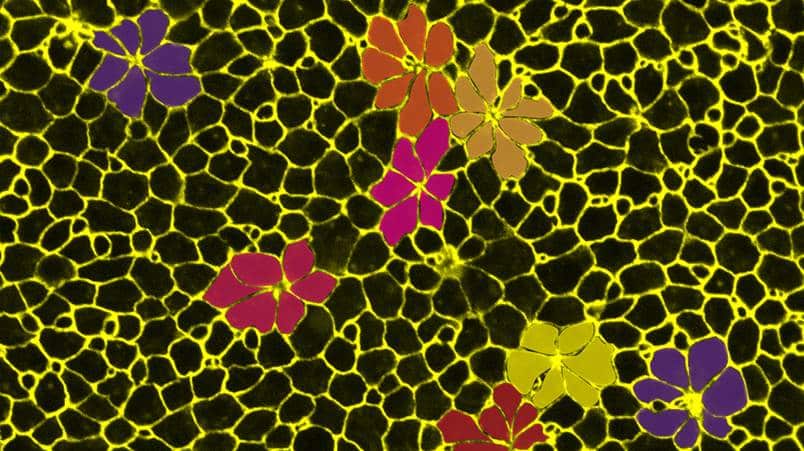
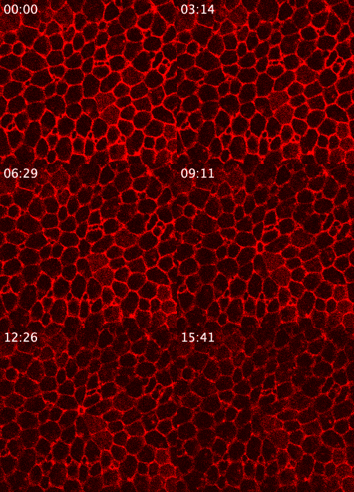
We therefore generated a CRISPR/Cas9 β-Catenin-mOrange2 knock in line, which has allowed us to label cell-cell adherens junctions and to visualise the dynamics of cell rearrangements and losses in the epidermis in vivo. Using confocal microscopy, we could follow the formation of rosette-like cell arrangements around cells that were being extruded from the epithelium.
The time-lapse videos of this process, which takes about 12 minutes from the initiation of rosette formation to complete extrusion, revealed that epithelial cell extrusion is a highly coordinated process in Nematostella. The central extruding cell becomes constricted, while the neighbouring cells form the characteristic flower-like rosette around the extruding cell. The continuity of cell-cell junctions in between rosette and extruding cells ensures the maintenance of epithelial integrity.
Once we had established the live-imaging approach, we moved beyond studying single extrusion events and developed a semi-automated image-analysis pipeline that allowed us to quantify cell area, cell density and rosette density across large datasets. Using this quantitative approach, we could ask how extrusion rates change across physiological states or after chemical inhibition of relevant signalling pathways, such as the nutrient-sensitive TOR pathway.
Moreover, we combined live imaging with fixed-tissue confocal microscopy and immunolabelling for F-actin, ERK signalling, apoptotic markers and TOR pathway activity. This combination of approaches allowed us to connect the dynamic cellular behaviour observed in vivo with its underlying molecular signalling. We found that the extruding cell undergoes apoptotic cell death while the surrounding rosette cells show active ERK signalling. Interestingly, we observed fragmented cell material within neighbouring cells, suggesting that material from extruded cells may be taken up and recycled. This was further supported by active nutrient-sensitive TOR signalling in late rosette cells.
Together, our findings in Nematostella show that both the phenomenology of the extrusion process and the underlying molecular mechanisms are highly conserved with those described in flies and vertebrates. This suggests that cell extrusion was likely already present in the common ancestor of cnidarians and bilaterians, where it could have served as a mechanism to remodel epithelia while maintaining tissue integrity and potentially to recycle cellular material in response to environmental stress.
Which imaging and image-analysis techniques did you use?
The β-Catenin knock-in line gave us a direct readout of epithelial cell boundaries in living animals. This was particularly important because the biological process we were interested in was fundamentally a change in cell geometry and cell-cell junctions.
We used confocal microscopy to image intact juvenile animals, rather that dissected epidermal tissue. The subcellular resolution allowed us to resolve the sequence of junctional rearrangements as neighbouring cells form a rosette around the extruding cell.
The second major component was quantitative image analysis. For this, we teamed up with bioinformatician PhD candidate Noah Bruderer and used Cellpose-based segmentation to identify individual epidermal cells and then developed a custom machine-learning workflow to ID rosettes. The pipeline allowed us to analyse images from many animals and quantify both cell area/density and rosette density across datasets.
What were the main technical challenges, and do you have any tips?
The biggest hurdle was simply keeping a juvenile Nematostella alive but sufficiently still to allow high-resolution imaging.
Juveniles are about 1x5mm in size, and thus much larger and more mobile than embryos. As they are literally water-filled epithelial sacks that can contract in all directions, they are notoriously difficult to immobilise. The challenge was to find the sweet spot where the animal is sedated enough to limit movements without perturbing cellular processes or even killing the animal.
We tested a number of approaches before settling on a relatively simple solution: 0.1M MgCl₂ in Nematostella medium combined with a new immobilisation setup. We repurposed multichannel Ibidi imaging slides, which are designed for cell culture and organoids, to gently squeeze and immobilise the polyp while allowing it to undergo confocal imaging.
This was probably one of the most important practical lessons from the project: the imaging setup can be just as important as the microscope.
The second major challenge was scaling the image analysis. Cell segmentation was not immediately automatic. We first had to manually segment and correct images to generate a suitable training dataset. We then had to manually identify and annotate rosettes to train the second machine-learning model.
We initially tried to identify rosettes primarily from geometric parameters, but this was not sufficiently robust. The cellular heterogeneity of the epidermis and the variability between rosettes made a more flexible machine-learning approach necessary.
Even after rosette prediction, we found that manual quality control was essential. In some cases, the model did not correctly identify every cell belonging to a rosette, so each detected rosette had to be checked and its constituent cells manually selected or corrected.
In the end, a human-in-the-loop approach was the most practical solution for us: manual annotation is used strategically to train the model, while automated prediction makes it possible to analyse large datasets that would otherwise be extremely time-consuming.
What advances in imaging or imaging analysis would help your research going forward?
The immediate next step is to make the analysis pipeline more automated. Reducing the amount of manual intervention during segmentation, rosette annotation and quality control would allow us to further accelerate the approach and apply it to much larger datasets.
We would also like to move from analysing individual images to analysing time-lapse video data directly. Our live imaging showed us the dynamics of rosette formation, but most of our large-scale quantitative analysis was performed on individual images. Automated cell tracking could allow us to characterise and quantify the entire extrusion process, including the timing and speed of each stage.
For larger-scale imaging, light-sheet microscopy could be particularly powerful for imaging entire juvenile animals. This could allow us to move beyond studying extrusion events within a local region of the epidermis and instead investigate cell behaviours across the whole animal during processes such as starvation-induced shrinkage. Combining whole-animal imaging with automated cell segmentation and rosette ID could ultimately provide a bridge between subcellular dynamics and organism-level physiological responses.
Another exciting direction would be to combine our β-Catenin transgenic line with additional reporters. For example, an ERK activity sensor could allow us to follow signalling dynamics during rosette formation in the same living animal. Combining structural dynamics with reporters for molecular signalling could help resolve the temporal sequence of both molecular and cellular events.
Ultimately, the goal is to combine high-resolution live imaging with scalable qualitative analysis. Developing imaging approaches that allow us to track cellular processes in whole, living animals opens up questions that are difficult to address using fixed sample alone.
For us, the most important step was simply being able to study the epidermis of living juvenile Nematostella polyps in such detail. Once we could do that, we could begin to connect cell behaviour, tissue biology and whole-animal physiology in a way that had not previously been possible in this system.
Hopefully, our work inspires other to tackle questions that require interdisciplinary approaches in non-model systems. For us, bringing together live imaging, quantitative image analysis and machine learning was essential and made the project a particularly enriching research journey.
Find the full story out now in Nature Communications.