

Imaging spotlight: linking mechanical forces and the Golgi
Posted by FocalPlane, on 22 September 2026
In this Imaging spotlight, Felix Campelo, Stéphanie Miserey and Jean-Baptiste Manneville highlight the key findings from their research asking whether the Golgi apparatus can sense and respond to mechanical forces. They talk about the importance of selecting the correct microscope for your question rather than assuming that the most sophisticated microscopy is best, and also discuss data representation and statistical analyses.
What are the key results from your paper?
We’ve known for more than a century that mechanical forces help shape cell and tissue function and development – it is worth looking back to D’Arcy Thompson’s 1917 book ‘On Growth and Form’, which is full of ideas connecting geometry, mechanics, and the biology of cells, tissues and organisms.
Cells in our bodies constantly experience a wide range of mechanical forces. They migrate across and adhere to extracellular matrices of different stiffness; airway cells experience forces as we breathe; and muscle cells are repeatedly stretched and compressed during contraction and relaxation. Cells can (and do) sense these forces (a process known as mechanosensing) through components of the plasma membrane, the main boundary between the cell and its environment, and also through the nucleus, the largest intracellular organelle. These mechanical inputs trigger signaling pathways (a process known as mechanotransduction) allowing cells to adapt their state and function to respond to their mechanical environment (a process known as mechanoresponse).
However, whether other intracellular structures can also sense and respond to extracellular mechanical cues has remained much less understood. So, we asked a simple question: does the Golgi apparatus sense and respond to mechanical forces?
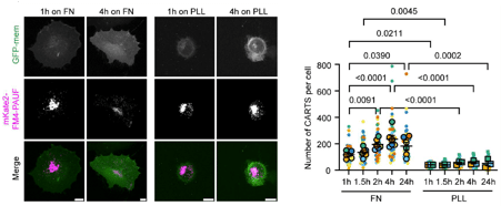
We found that, indeed, it does. Using several complementary mechanical perturbations (including cell spreading, substrates of different stiffness, and direct mechanical stretching), we found that extracellular mechanical cues regulate the formation of secretory carriers at the Golgi apparatus (Fig. 1). Cells that spread efficiently and formed robust focal adhesions produced more Golgi-derived carriers. Cells grown on stiffer substrates also showed a comparable increase, and directly stretching cells similarly stimulated secretory carrier biogenesis.
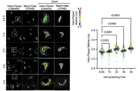
Next, using the membrane tension reporter Halo-Flipper together with fluorescence lifetime imaging microscopy (FLIM), we found that extracellular mechanical forces had also an impact on the biophysical properties of Golgi membranes. During cell spreading, for example, the fluorescence lifetime of Halo-Flipper at the Golgi apparatus progressively increased, consistent with an increase in membrane tension and/or lipid packing (Fig. 2). Similarly, cells grown on soft substrates showed lower Golgi Halo-Flipper lifetimes than cells grown on glass.
We also identified components of the pathway linking these mechanical inputs to Golgi function. Cell spreading increased microtubule acetylation, and pharmacologically increasing microtubule acetylation also, in turn, enhanced Golgi export. Mechanical cues were also associated with changes in the levels of the lipid diacylglycerol (DAG) and in protein kinase D (PKD) activity at the Golgi apparatus. This is relevant because both DAG and PKD are important regulators of the biogenesis of post-Golgi carriers, such as CARTS (carriers of the TGN to the cell surface).
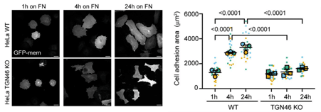
Perhaps the result we find most interesting from a conceptual point of view is that this communication works in both directions. When we inhibited Golgi export, either pharmacologically or genetically, cells could no longer spread normally (Fig. 3). We therefore propose a reciprocal feedback loop: extracellular mechanics regulate Golgi function, and Golgi-derived trafficking, in turn, helps cells adhere and adapt to their immediate environment. This places the Golgi apparatus as an active participant in cellular mechanoadaptation and not simply as a downstream secretory factory.
Which imaging/image analysis techniques have you used?
We used a combination of imaging approaches because the different questions in the study required quite different kinds of information.
For live-cell imaging of secretory carriers arriving at the plasma membrane, we used total internal reflection fluorescence (TIRF) microscopy. This was particularly useful for visualising CARTS containing the specific secretory cargo protein PAUF (pancreatic adenocarcinoma upregulated factor) close to the basal plasma membrane and asking where at the plasma membrane they are delivered. Because TIRF restricts excitation to a very thin region adjacent to the coverslip to which cells adhere, it provides a much cleaner view of membrane-proximal carrier dynamics than conventional widefield or confocal imaging. We could therefore track the movement of individual carriers as they approached towards focal adhesion-rich regions of the plasma membrane, where they paused for some seconds after which the fluorescence vanished, consistent with an event of carrier fusion with the plasma membrane.
For most fixed-cell imaging, we used confocal or spinning-disk confocal microscopy. These experiments required imaging of the whole cell volume, e.g., to count the number of post-Golgi carriers per cell, examine microtubule acetylation, or measure Golgi-localized DAG levels and PKD activity. We also used widefield fluorescence microscopy for experiments involving the equibiaxial mechanical stretching device. Here, the priority was robust imaging of cells that were seeded on the stretchable PDMS membrane and under mechanical strain, rather than optical sectioning or enhanced spatial resolution. The relatively large thickness of the PDMS membrane also imposed an important geometrical constraint, so we needed to use an upright microscope, instead of the more usual inverted microscope configuration, allowing us to image the cells “directly” without the excitation and emitted light having to travel through the thick PDMS layer.
A particularly important technique we used for this work was fluorescence lifetime imaging microscopy (FLIM), which we combined with the Halo-Flipper membrane probe. Halo-Flipper is a modified variant of the Flipper family of membrane probes that acts as a ligand for HaloTag. Flipper responds to changes in the mechanical state of the membrane, including lipid packing and membrane tension, which can be detected by the concomitant change in its fluorescence lifetime. We targeted Halo-Flipper to the Golgi membranes using a Golgi-localized HaloTag protein. Because some Halo-Flipper signal was also present outside the perinuclear Golgi area, we needed to label the Golgi-localized HaloTag protein in parallel with a spectrally different far-red HaloTag ligand (JF646 in our case). With this double labeling, we were able, during image analysis, to define a Golgi mask and restrict Halo-Flipper lifetime measurements to that region (Fig. 2). This gave us a spatially specific readout of how extracellular mechanical forces affect the mechanical properties of the Golgi membranes in living cells.
Are there any technical tips or tricks that you have learnt while doing this research?
After working with microscopy for many years, something that we believe is quite important to take into account is that the most sophisticated microscope or the one with the highest resolution is not necessarily the best microscope for every experiment. The first questions should always be: What information do we actually want to obtain? Will the experiment be in fixed or in living cells? What spatial and temporal resolution are required? If you are imaging living cells, is it fast or long-term imaging that you need? How much signal is available, how much illumination can the sample tolerate before going funky? And so on, so forth. Microscopy, like life, involves compromises. Compromises between spatial resolution, temporal resolution, signal-to-noise ratio, phototoxicity, etc. Improving one parameter often comes at the expense of another, so choosing the right technique is really about matching these trade-offs to the biological question rather than simply using the highest-resolution instrument available. In our case, this also meant accepting practical constraints imposed by the experimental setup (such as the geometry of the mechanical stretching device, and the inherent photophysical properties of Flipper dyes) and designing the optimal imaging of our purposes. Finally, for reliable and consistent quantitative fluorescence imaging, another apparently simple but extremely important point is to avoid detector saturation and keep identical acquisition settings between conditions.
We also aimed to clearly and openly distinguish between individual cell measurements (different cells being measured per condition within a single experiment) and reproducibility between independent biological replicates. For this reason, we used SuperPlots for both data representation and statistical analyses (see right panels in Figs. 1–3). SuperPlots show the distribution of measurements from individual cells and differentiate which cells belong to each of the biological replicates. Statistical comparisons were then based on a central (e.g., mean or median) value of the distribution measured for each biological replicates rather than treating every cell pooled across experiments as an independent observation.
Working with Halo-Flipper and FLIM required us to be particularly careful. One important practical point here is to minimize sample illumination as much as possible before the actual FLIM acquisition. Flipper probes can act as photosensitisers, so excessive exposure to light can cause changes in the membranes you want to measure. Spatial specificity of the Halo-Flipper signal was another important consideration. Because some of the signal can appear to be unspecific, we found it essential to define the Golgi area independently and restrict lifetime analysis to that region. For that, given the relatively broad emission spectrum of Flipper, we needed to use a spectrally well-separated far-red marker (JF646 in our case). We also found it quite useful and reassuring to include a perturbation for which the expected direction of the mechanical change had already been measured by other means. Latrunculin A, a chemical that perturbs the actin cytoskeleton and had been previously shown to reduce Golgi rigidity in mechanical measurements, produced the expected decrease in Halo-Flipper lifetime. This provided an important internal control and increased our confidence that the probe was sensitive to changes in the biophysical state of Golgi membranes in our experimental system.
Are there any advances in imaging or image analysis that would help your research?
A major goal for the field is to move from relatively global mechanical measurements towards more local and spatially resolved force maps. The Golgi apparatus is a highly dynamic organelle with a quite complex three-dimensional structure, and we still know very little about how its membranes are laterally organized, and even less about how forces are distributed, generated and transmitted along their surface. Imaging tools that could measure and/or manipulate membrane forces locally, with high spatial and temporal resolution, would therefore be extremely valuable.
We would also benefit greatly from more sensitive Golgi-targeted probes for membrane mechanics. Halo-Flipper has been very useful, but because its fluorescence lifetime changes not only with membrane tension but also with lipid packing, having orthogonal tools that provide independent mechanical measurements would help considerably.
Another important advance would be to have improved live-cell reporters for Golgi lipids and signaling activities. Ideally, we would like to simultaneously follow mechanical changes, DAG levels, PKD activation and secretory carrier formation at the Golgi membranes in individual living cells. This would allow us to establish, in a much more precise manner, the spatiotemporal sequence of the events leading to transport carrier biogenesis (from cargo sorting and loading to membrane budding and fission) and determine how each of these steps is influenced by mechanical stimuli.