

Featured image with Shang-Wu Shih
Posted by FocalPlane, on 9 October 2026
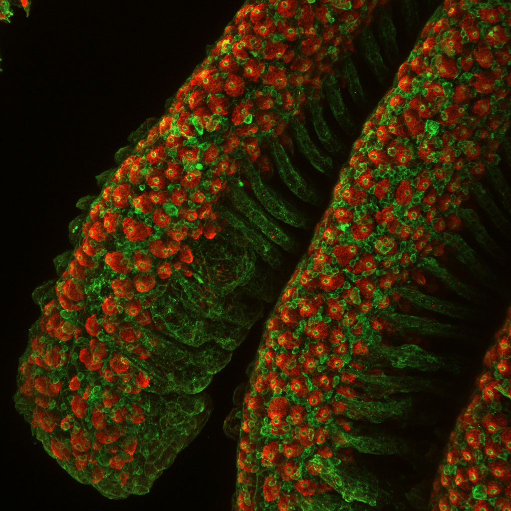
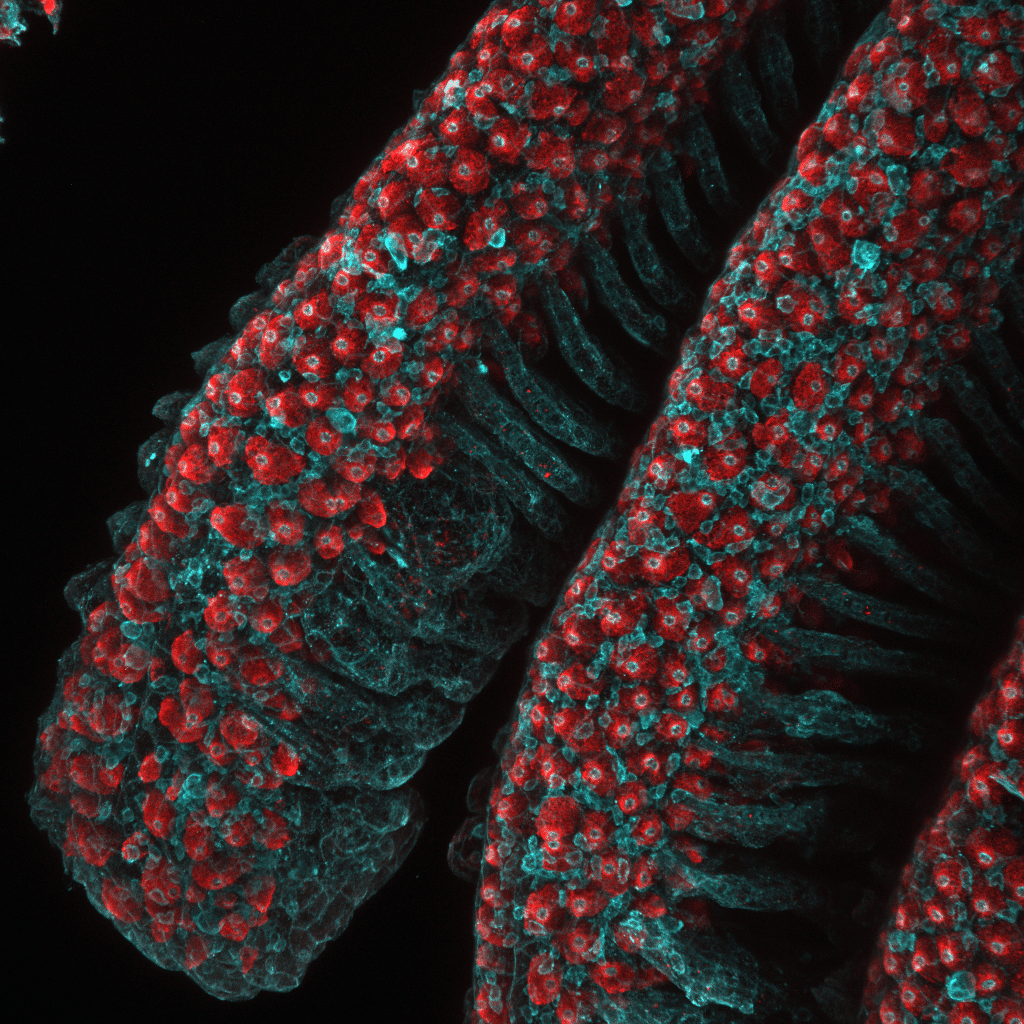
Our featured image, acquired by Shang-Wu Shih, shows ionocytes distributed along the gill filaments of marine medaka. These specialized epithelial cells are responsible for maintaining systemic ion and acid–base homeostasis. Gill tissues were fixed and immunostained for Na⁺/K⁺-ATPase (NKA, red), a marker of ionocytes, and actin (green/cyan), which outlines the tissue architecture. Images were acquired using a ZEISS LSM 980 confocal microscope and processed in Fiji as maximum-intensity Z-stack projections.
Read more about Shang-Wu’s research.
Research career so far: I completed my PhD at National Taiwan University (NTU) in Taiwan, where I studied fish physiology, particularly the biology of ionocytes. I am currently a postdoctoral researcher at the University of Michigan. Throughout my research, I have been fascinated by how epithelial cells sense environmental changes and maintain tissue function, and advanced microscopy has been an essential tool for answering these questions.
Current research: My current research investigates how fully differentiated epithelial cells maintain tissue homeostasis under environmental stress. Using zebrafish ionocytes as a model, I study how calcium signaling controls the transition between cellular quiescence and proliferation. Ultimately, I hope this work will reveal conserved mechanisms of epithelial plasticity that are relevant across vertebrates.
Favourite imaging technique/microscope: Confocal microscopy is my favorite imaging technique because it allows me to visualize protein localization and cellular organization with excellent resolution. The ZEISS LSM 980 with Airyscan has become my preferred microscope, as it combines high sensitivity with enhanced spatial resolution, making it especially valuable for imaging epithelial tissues.
What are you most excited about in microscopy? I am most excited by the ability to image living tissues over time with increasingly high spatial and temporal resolution. Watching individual cells change their behavior in real time, rather than relying on static snapshots, is transforming how we study tissue homeostasis and regeneration. I believe future advances that combine live imaging with genetic tools and single-cell technologies will provide unprecedented insights into cell behavior in vivo.