

Behind the paper: How tensin 1 condensates regulate cell adhesion dynamics
Posted by FocalPlane, on 27 October 2026
Posted on behalf of Michal Dibus
Adhesions, condensates, and tensin 1
One of our many research interests in the Ivaska lab is understanding how cells interact with the extracellular matrix (ECM) to sense and respond to biochemical and mechanical cues from their environment. In particular, we are interested in integrin-mediated cell adhesions, which connect the ECM to the cell cytoskeleton through a multimolecular complex of adhesion scaffold and adaptor proteins [1]. One such protein is tensin 1 (TNS1), a multidomain adaptor protein that directly links active integrins to the actin cytoskeleton, and the main protagonist of our recent story [2].
To facilitate cell migration, integrin adhesions must be highly dynamic, undergoing continuous assembly, maturation, and disassembly. In recent years, these dynamics have been linked to the ability of several adhesion proteins to undergo phase separation and form membraneless condensates. While investigating the biological roles of tensins in cell adhesion, we observed that TNS1 forms cytoplasmic condensates when overexpressed in cells. This prompted us to investigate the role of TNS1 phase separation in regulating adhesion maturation and dynamics.
We first confirmed that TNS1 does indeed forms condensates using a variety of imaging approaches including live-cell imaging and correlative light-electron microscopy (CLEM), which allowed us to demonstrate that TNS1 condensates are highly dynamic membraneless structures. Although the overexpression system used in our initial experiments provided valuable insights into the properties of TNS1 condensates, it did not reveal how TNS1 behaves at endogenous expression levels. We therefore generated a cell line with endogenously tagged TNS1 and demonstrated that endogenous TNS1 condensates form in response to limited integrin ligand availability (Figure 1) or following acute adhesion disassembly induced by the loss of actomyosin contractility.
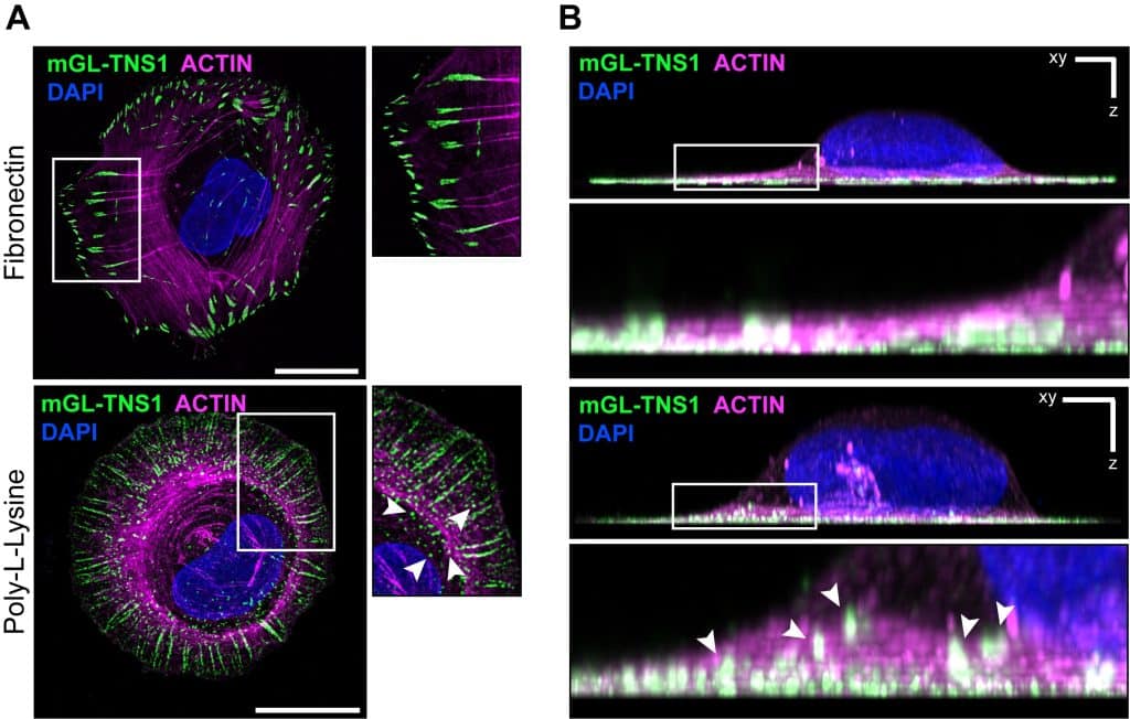
We further employed proximity-dependent biotinylation proteomics (BioID) to identify the main components of TNS1 condensates. Although this approach allowed us to identify a condensate-enriched TNS1 proximity interactome, extensive validation was needed to distinguish genuine condensate components from proteins associated with TNS1 at other cellular locations, such as focal adhesions and membrane ruffles. Ultimately, we demonstrated that TNS1 condensates selectively recruit several adhesion proteins, including TNS3, zyxin, paxillin, p130Cas and others, predominantly in their unphosphorylated and thus largely inactive states.
The elephant in the room: what do the TNS1 condensates do?
One of the major challenges in the phase separation field is to elucidate the physiological functions of biomolecular condensates [3]. Our observations that endogenous TNS1 condensates form upon acute adhesion disassembly or under conditions of limited integrin-ECM engagement led us to hypothesize that they might serve as temporary reservoirs of unused adhesion proteins. This hypothesis was further supported by our finding that TNS1 condensates selectively recruit unphosphorylated adhesion proteins.
We decided to test this from two complementary perspectives.
First, to investigate the potential role of TNS1 condensates in storing adhesion proteins, we acutely induced condensate formation and observed an increase in the expression of adhesion proteins recruited to the condensates. In contrast, the expression levels of adhesion components that were not recruited to TNS1 condensates remained unchanged. These findings suggested that TNS1 condensates can stabilize the expression levels of recruited proteins, potentially by protecting them from degradation.
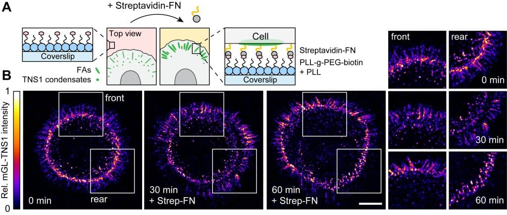
Second, we wanted to investigate whether proteins stored in TNS1 condensates could be made available for the formation of new adhesions. Capturing the dynamic interplay between condensates and adhesions proved technically challenging, mostly due to the highly dynamic nature of the condensates, rapid adhesion turnover, and photobleaching of the endogenously tagged TNS1 during live-cell imaging. We therefore adapted our dynamic micropatterning approach [4] to rapidly induce adhesion formation in cells seeded on surfaces with limited ECM availability. By combining this approach with live-cell imaging, we showed that upon increase in integrin-ECM engagement some of the condensates close to the cell periphery disassembled, correlating with the increased formation of new TNS1-positive adhesions in their proximity (Figure 2). We further supported these findings using live-cell imaging of cells seeded on a surface with limited integrin ligand availability (poly-L-lysine), in which we induced integrin activation and clustering by adding manganese ions (Video 1). These observations support our model that condensate disassembly increases the local availability of adhesion components, facilitating their recruitment into newly forming adhesions.
Keeping the condensates in check: protein disorder and phosphorylation
Having established the biological functions of TNS1 condensates, several important questions remained: What drives their assembly and disassembly? How is this process regulated? And how does it influence adhesion dynamics and cell behaviour?
Protein condensation is generally driven by multivalent interactions involving structured protein domains, weak interactions mediated by intrinsically disordered regions (IDRs), or a combination of both [5]. Using a series of carefully designed TNS1 deletion mutants, we demonstrated that condensate formation is mediated by its IDR, particularly its C-terminal part. We further showed that the amino acid sequence of the IDR is a major determinant of condensate properties. Importantly, cells expressing a TNS1 mutant lacking the IDR, which was unable to form condensates, exhibited increased adhesion dynamics accompanied by enhanced cell migration and invasion compared with cells expressing wild-type (WT) TNS1. These findings suggest an important role for the TNS1 IDR in regulating adhesion stability and, consequently, cell behaviour.
Since phosphorylation has been shown to regulate condensate formation in a number of proteins, we hypothesized that the same might be true for TNS1, particularly as a previous study had demonstrated extensive serine and threonine phosphorylation of TNS1 in response to cellular stress [6]. Indeed, we found that activation of stress-responsive kinases promoted TNS1 phosphorylation and triggered condensate disassembly (Video 2). Consistent with our IDR deletion experiments, a phosphomimetic TNS1 mutant that was unable to form condensates also exhibited increased adhesion dynamics and cell migration.
Overall, our findings reveal phosphorylation as a key regulator of TNS1 condensation and suggest that this mechanism allows cells to dynamically control the storage and availability of adhesion proteins, thereby influencing adhesion dynamics and cell migration.
Finally, beyond its contributions to cell biology, our work might also be of particular interest to microscopy enthusiasts. In this multidisciplinary study, we combined immunocytochemistry, live-cell imaging with high temporal resolution, fluorescence recovery after photobleaching (FRAP), CLEM, and superresolution microscopy to investigate the biological functions of TNS1 condensates. In addition, we adapted our dynamic micropatterning approach [4] to acutely induce focal adhesion assembly, allowing us to explore the dynamic crosstalk between TNS1 condensates and newly forming adhesions. Importantly, we also used cells expressing endogenously tagged TNS1 to validate our observations from overexpression systems. Together, we believe these approaches provide a valuable toolkit for studying phase separation of adhesion proteins and its role in regulating adhesion dynamics.
The full story is available at https://doi.org/10.1038/s41467-026-74001-3.
References
1. Chastney, M.R. et al. (2025) The role and regulation of integrins in cell migration and invasion. Nat. Rev. Mol. Cell Biol. 26, 147–167
2. Dibus, M. et al. (2026) Adhesion-derived condensates control component availability to regulate adhesion dynamics. Nat. Commun. 17, 7222
3. Aierken, D. et al. (2026) Roadmap for Condensates in Cell Biology. PRX Life 4, 032501
4. Isomursu, A. et al. (2023) Dynamic Micropatterning Reveals Substrate‐Dependent Differences in the Geometric Control of Cell Polarization and Migration. Small Methods 2300719, 1–17
5. Musacchio, A. (2022) On the role of phase separation in the biogenesis of membraneless compartments. EMBO J. 41, 1–20
6. Hall, E.H. et al. (2010) Comprehensive analysis of phosphorylation sites in tensin1 reveals regulation by p38MAPK. Mol. Cell. Proteomics 9, 2853–2863
Get involved
Create an account or log in to post your story on FocalPlane.
More posts like this

Filter by
- NewsApply
- DiscussionsApply
- How toApply
- ToolsApply
- Case studiesApply
- InterviewsApply
- JobsApply
- EducationApply
- Blog seriesApply
- Latin American Micro..scopistsApply
- Bio-image Analysis w..ith NapariApply
- Imaging with…Apply
- Towards Global Acces..sApply
- Latin America Bioima..gingApply
- From Zero to Qupath ..HeroApply
- Asian Microscopists ..and Cell BiologistsApply
- AIC at HHMI JaneliaApply
- Deep Learning for Bi..o-image analysisApply
- GloBIAS – updates fr..om the communityApply
- WAMBIAN: West Africa.. in FocusApply
- Volume EMApply
- Highlights from Euro..-BioImagingApply
- LSFM seriesApply
- DIY MicroscopyApply
- View all