Advertisement

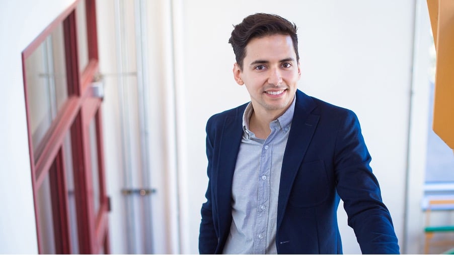
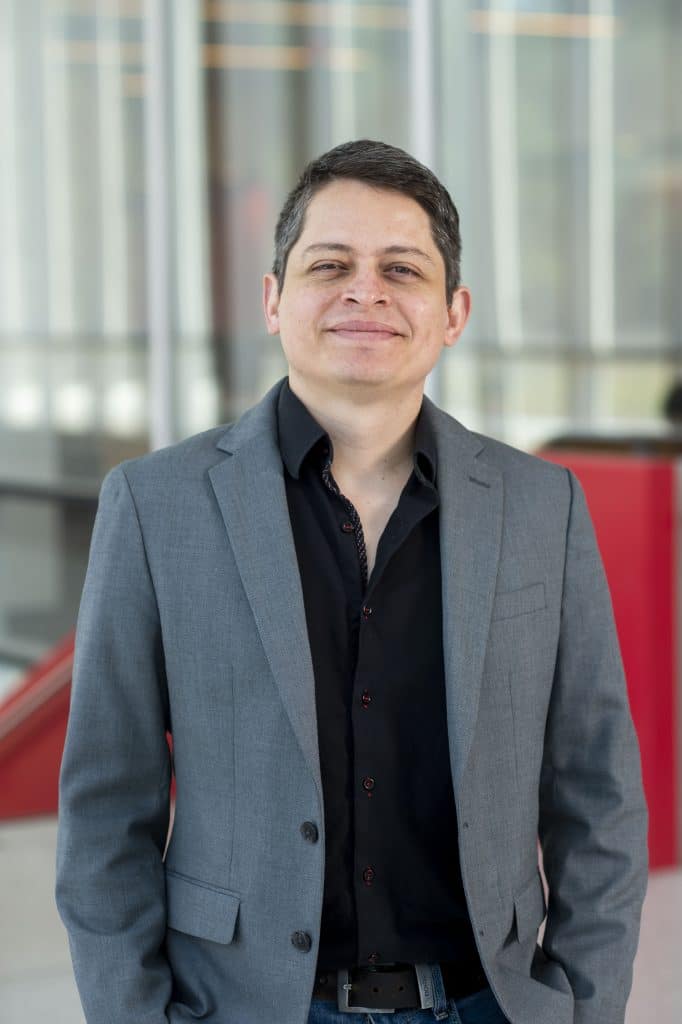
An interview with Sebastian Lourido

Posted by , on 21 November 2023

MiniBio: Prof. Sebastian Lourido is a tenured professor at the Whitehead Institute and the Faculty of Biology at MIT in Boston, USA. He began his studies in Colombia, and from an early age developed an interest for the natural world. Seeking versatility in his undergraduate degree, he went to Tulane, in the USA, where he

Microscopy preprints – bioimage analysis tools

Posted by , on 17 November 2023

Here is a curated selection of preprints published recently. In this post, we focus specifically on new tools for bioimage analysis and data management.

Featured image with Nick Gatford

Posted by , on 16 November 2023

Our featured image shows a single human dopaminergic neuron generated from a human stem cell, acquired via super-resolution Airyscan confocal microscopy at the University of Oxford Micron facility by Nick Gatford. We caught up with Nick to find out about his research and what he is excited about in microscopy.

Contribute to Euro-BioImaging’s growth!

Posted by , on 15 November 2023

The Euro-BioImaging Hub team is growing fast thanks to EU project funding. Come and join our outstanding team and shape the future of European biological and biomedical imaging!

Featured image with Oona Paavolainen

Posted by , on 15 November 2023

Our featured image, acquired by Oona Paavolainen, shows a primary mammary gland organoid, in a collagen-I matrix, formed from a single primary epithelial cell isolated from material obtained from a breast reduction surgery. The image was taken with a spinning disk confocal microscope (3i CSU-W1 Spinning disk) using a 20x Zeiss Plan-Apochromat (NA=0.8) objective. The

How to Minimise Photodamage with LED illumination

Sponsored by CoolLED, on 15 November 2023

Both fixed and live samples are susceptible to photodamage during fluorescence microscopy, which can shorten time-lapse experiments and cast doubt on observations. LED illumination for widefield fluorescence has the potential to counter this serious problem, whether running a complete experiment or simply finding the region of interest prior to confocal microscopy. Exposing biological samples to

Featured image with Till Stephan

Posted by , on 14 November 2023

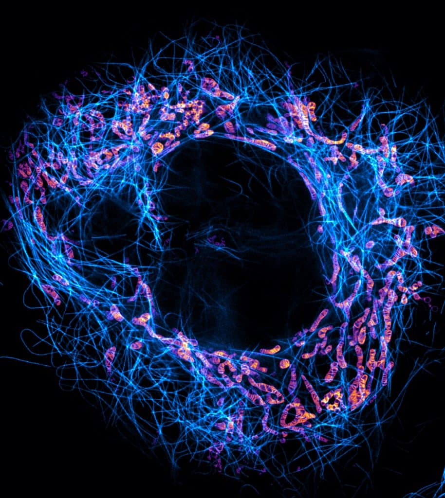
Our featured image, acquired by Till Stephan, shows a live-cell stimulated emission depletion (STED) microscopy recording of a HeLa cell in which mitochondria (orange-magenta) and microtubules (cyan) are labelled. The super-resolution image reveals the densely stacked mitochondrial cristae, coordinated invaginations of the mitochondrial inner membrane.

An interview with Iván Rey

Posted by , on 14 November 2023

MiniBio: Iván Rey Suarez is a postdoctoral associate at a joined position at Universidad de los Andes in Colombia, and the Institute for Physical Science and Technology (IPST) at the University of Maryland, where he focuses on studying the mechanobiology of the cytoskeleton. Iván developed a passion for nature and for physics since an early

FocalPlane playlist: Christophe Leterrier

Posted by , on 14 November 2023

Discover the preprints, papers and FocalPlane content that Christophe Leterrier has found interesting in recent months!

New look for FocalPlane

Posted by , on 13 November 2023

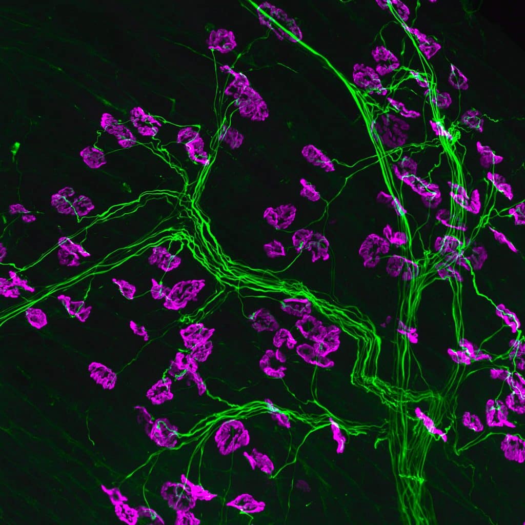
We are delighted to launch a new look for FocalPlane. Every day this week, we’ll be releasing a new banner image and highlighting the scientist that acquired it. Today, we start with an image from Rebecca Simkin. Rebecca won our 2023 image competition, and you can read our featured image post with Rebecca here: https://focalplane.biologists.com/2023/09/01/and-the-winner-is/