Advertisement

How To Train an Undergraduate Researcher in The Age of COVID-19

Posted by , on 10 August 2020

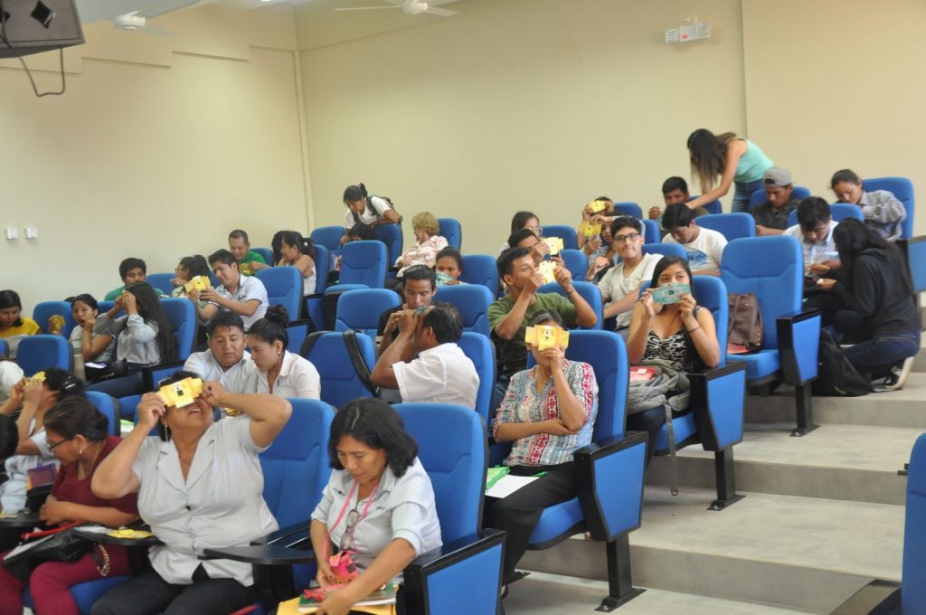
The year 2020 has been challenging for researchers around the world due to the COVID-19 pandemic. With limited access to our labs, it is not easy to gain hands-on bench experience. We are the undergraduate researchers in the Rodal Lab, from Brandeis University in Waltham, MA. Our lab primarily studies membrane trafficking events at the

Science and Art – the not so odd couple?

Posted by , on 31 July 2020

At a first, superficial glance, you could be forgiven for placing scientists and artists at the opposite ends of the career spectrum. Scientists need to be accurate and methodical. They must generate highly reproducible data while adhering to strict regulations. On the other hand, artists are often stereotyped as disorganized, free spirits, ungoverned by rules,

Expansion microscopy

Posted by , on 29 July 2020

Written by Shoh Asano and Ruixuan Gao Light microscopy and diffraction limit For centuries, light microscopy has played a central role in biological studies. The first implementations of a light microscope dates back to as early as the late 16th and early 17th century, when an array of polished lenses was used to magnify (biological)

“openFrame” for modular, extensible, easily maintained, open-source microscopy

Posted by , on 23 July 2020

The openFrame [1] is an open-source microscopy hardware project initiated by the Photonics Group in the Physics Department at Imperial College London that is intended to provide access to a modular, upgradable, easily maintained and modifiable microscope frame that can be implemented at relatively low cost compared to existing commercial instruments. openFrame is a component

Electron microscopy: from the dark ages to a bright future

Posted by , on 21 July 2020

Good sample preparation is, as every microscopist knows, the key to delivering sound results from an imaging experiment. In the digital age, and with the advent of big data, image analysis is also critical to extract meaningful quantitative results from image data. Indeed nowadays, the microscope itself is often the most well-developed and user-friendly part

Take this quick X-Ray Microscopy Usage Survey!

Posted by , on 17 July 2020

Take part in online poll to gauge use of technique and identify opportunities to expand its application The use of X-ray microscopy techniques is well established in many scientific and engineering disciplines but not all have “seen the light”. The X-ray Microscopy Focussed Interest Group (X-ray FIG) of the Royal Microscopical Society (RMS) would like

Etch A Cell - segmenting electron microscopy data with the power of the crowd

Posted by , on 16 July 2020

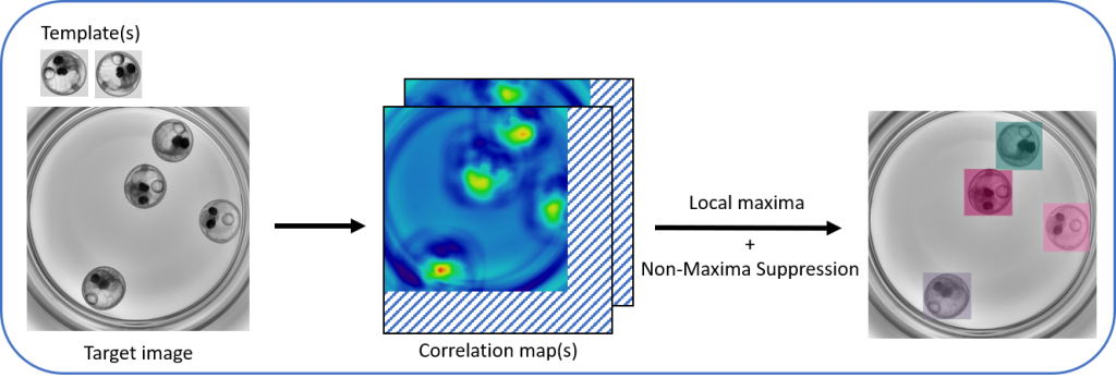
Recent years have seen remarkable developments in imaging techniques and technologies, producing increasingly rich datasets that require huge amounts of costly technological infrastructure, computational power and researcher effort to process. Techniques such as light-sheet microscopy and volume electron microscopy routinely generate terabytes worth of data overnight. With a single data acquisition producing more images than

Primers on Microscopy for Biologists - Resolution

Posted by , on 15 July 2020

Formal definitions of resolution refer to imaginary objects such as infinitely small sources of light. I will avoid those and instead try to provide a pragmatic explanation. Practically, the spatial resolution is the size of the smallest structure that can be distinguished in the light coming from a specimen. All sensors and components of the

How CLIJ2 can make your bio-image analysis workflows incredibly fast

Posted by , on 14 July 2020

Do you also spend a substantial amount of your time waiting for automated image analysis to finish? I did, again and again for more than a decade. And, then,  the morning after running a script overnight, you realize that a parameter was wrongly set. It was about time to change that. History Processing on graphics

Microscope technology development and the search for a free lunch

Posted by , on 10 July 2020

I fortuitously stumbled into the field of microscopy around 2013, just in time to enjoy the hype of the 2014 Chemistry Nobel Prize for super-resolved fluorescence imaging. As a newcomer, I was quite surprised that this nearly 400-year-old field could still catch the attention of Stockholm. After all, wasn’t a microscope just some lenses to