/* */

Advertisement

Displaying posts with the tag: is_archive

Tracking in napari

Posted by , on 1 June 2023

In this blog post we are exploring cell tracking in napari. Two important processes in normal tissue development and disease are cell migration and proliferation. To gain a better understanding on these processes, tracking in time-lapse datasets is needed. By measuring track properties, like velocity and the total travelled distance, spatio-temporal relationships can be studied.

Feature extraction in napari

Posted by , on 3 May 2023

This blog post revolves around extracting and selecting features from segmented images. We will define the terms feature extraction and selection. Also, we will learn how to categorize features and can look up specific features in a glossary. Furthermore, we will explore how to extract features in napari. Definition of feature extraction During feature extraction,

Quality assurance of segmentation results

Posted by , on 13 April 2023

This blog post revolves around determining and improving the quality of segmentation results. A common problem is that this step is often omitted and done rather by the appearance of the image segmentation than by actually quantifying it. This blogpost aims to show different ways to achieve this quantification as this leads to reproducibility. Therefore,

Annotating 3D images in napari

Posted by , on 30 March 2023

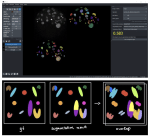
This blog post revolves around generating ground truth in 3D images for segmentation. Therefore, we will define what ground truth is and how we can generate it using napari in a time-efficient way. We will also learn about difficulties of annotating alone or in groups and address possible solutions for both. Challenges of image segmentation

Rescaling images and pixel (an)isotropy

Posted by , on 2 March 2023

This blog post shows the importance of rescaling 3D image data and what anisotropy of pixels/voxels has to do with it. We will see which different options exist to rescale an image as well as their advantages and limitations. We will also apply a commonly used segmentation algorithm, Voronoi-Otsu-Labeling, on the original and the rescaled

Explorative image data science with napari

Posted by , on 23 May 2022

When analysing microscopy image data of biological systems, a major bottleneck is to identify image-based features that describe the phenotype we observe. For example when characterising phenotypes of nuclei in 2D images, often questions come up such as “Shall we use circularity, solidity, extend, elongation, aspect radio, roundness or Feret’s diameter to describe the shape