

Imaging spotlight: fluorogenic L-DNA probes
Posted by FocalPlane, on 6 August 2026
In this Imaging spotlight, we hear from Bas van Bommel and Helge Ewers about the fluorogenic L-DNA probes they used to optimise DNA-PAINT labelling for single molecule localisation microscopy.
Can you briefly describe DNA-PAINT, and the advantages of your L-DNA probes?
DNA-PAINT is a super-resolution microscopy method developed by Ralf Jungmann (Jungmann et al., Nature Methods 2014) that like STORM and PALM is based on the highly accurate localization of many, many individual molecules and the rendering of an image from the thousands of localizations. DNA-PAINT uses the kinetically very well-understood transient binding of short DNA-oligomers to their reverse complement to deliver a dye to a target structure via a DNA-coupled antibody or other binder. Since many sequences are available even for the typically used nonamer oligo, it can be used for multiplexed imaging of many targets, which makes it special. Also, since it does not rely on blinking dyes, but allows researchers to pump many photons through very bright and stable fluorophores, usually the reached accuracy is higher.
Now the problem with DNA is, it is of course also present in the cell, especially in the nucleus. A typical nonamer for example will statistically have tens of thousands of binding sites in a diploid human genome. As a result, there will be background binding of dye-coupled oligos. That is of course a problem in a single molecule technique, as a single localization is a single localization, no matter if “real” or resulting from background binding.
When postdoc Hylkje Geertsema joined my lab, she decided to ask, if this could be circumvented by using left-handed DNA (L-DNA) for DNA-PAINT. L-DNA is chemically identical to the right-handed DNA in life, but its opposite in chirality or structural handedness. This L-DNA cannot interact with right handed DNA at all. When she used it in cells, she found that it reduced background in DNA-PAINT imaging of the nucleus significantly (Geertsema et al., Nature Biotechnology 2021). This will of course be of great advantage in DNA-PAINT investigation of nuclear structures, chromosomes and so on and allow multiplexed imaging like DNA-PAINT.
What have you labelled with your L-DNA probes? Do you foresee there being any difficulties extending this to other targets or models?
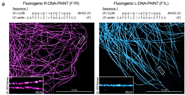
As super-resolution people, our first proof-of-principle target is always microtubules in cultured cells. And there, L-DNA performed absolutely identically to R-DNA. After this, we of course went for nuclear structures, so we labeled Ki67, and PCNA, prominent proliferation markers and replication forks labeled via BrdU. We also performed immuno DNA-FISH experiments. We performed the experiments with R-DNA and L-DNA one after the other in the same sample by exchanging probes to exclude cell-to-cell variation error. In all cases we had less background when we imaged with L-DNA than with R-DNA, regardless which probe we used first.
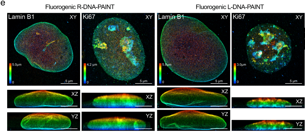
Bas van Bommel, a postdoctoral fellow in my lab now combined L-DNA-PAINT with an idea from Joerg Bewersdorf, the use of fluorogenic oligomers that are only fluorescent when bound to the target (Chung et al., Nature Methods, 2022). This makes 3D imaging in DNA-PAINT much easier and the entire DNA-PAINT procedure faster. Bas added beautiful 3D images of the nuclear envelope protein laminate and again imaged Ki67, in 3D now. In his recent paper we discuss here (van Bommel and Ewers, Nano Letters 2026) he also characterized the fluorogenic L-DNA probe intensively and provides a lot of very useful background information. In theory, this should be useful in tissue, organoids or other samples as the fluorogenic nature allows for 3D imaging.
Naïve question: can you just ask companies to synthesise L-handed DNA oligos?
That is indeed a good question, not many companies make L-DNA actually, so you have to look around a bit, but a simple search will find you a provider. Surprisingly it is not much more expensive than R-DNA. There are actually no tricks, that is the magic of chiral chemistry. Kinetics, labeling reactions, affinity are all the same. So just do what you do for right-handed DNA and it will work.
What are the prospects for further development?
Technically, everything that works for R-DNA could be done with L-DNA and thus looking at the DNA-PAINT world, the possibilities to use L-DNA are endless. One thing is using L-DNA to expand the portfolio of available oligomers for multiplexed imaging and Ralf Jungman went into that direction (Unterauer et al., Nature Communications 2025). Probe designs containing PRG-spacers can further improve binding kinetics as young PI Florian Schueder has shown https://www.biorxiv.org/content/10.64898/2026.03.23.710523v1; these may likewise be used with L-DNA.
Technically, L-DNA could also be used in other imaging modes than DNA-PAINT such as confocal imaging or STED by changing oligomer length so that the dye stays on the target for longer. This should be useful. For any investigation of DNA-rich structures at the nanoscale our approach should make a difference. We are looking for interested collaborators or postdoctoral fellows that work on questions regarding 3D nanoscale organization in the nucleus, so get in touch if you are interested.
Where can people find more information?
The papers cited above contain a lot of information to get you started for experiments. Besides this, we are happy to help if there are any specific questions.