

Seeing comes before knowing: how live imaging led to the discovery of blood vessel-resident macrophages
Posted by FocalPlane, on 12 August 2026
by Bart Weijts and Catherine Robin
Our laboratory is driven by one fundamental question in developmental biology: how hematopoietic stem and progenitor cells (HSPCs), which establish lifelong blood production, are generated and expand during development, and how these processes are regulated. Answering this question is important not only for understanding blood development but also for advancing regenerative medicine. HSC transplantation remains the only curative treatment for many hematological disorders, yet finding compatible donor cells is often challenging. Despite considerable efforts, generating functional HSCs de novo in the laboratory remains remarkably inefficient. Understanding how HSCs are naturally produced during embryonic development is therefore essential for designing new strategies to generate transplantable HSCs.
To study HSPC development, we exploit one of the greatest advantages of the zebrafish embryo: its optical transparency. Live imaging allows us to directly visualize HSPCs in real time and follow their emergence, migration, colonization, and interactions with their developmental niches in vivo at single-cell resolution. Over the years, we have learned that patiently watching embryos often reveals biology that would otherwise remain hidden, sometimes sending a project in an entirely new direction.
While analyzing time-lapse recordings of the HSPC developmental journey, we repeatedly noticed an unusual population of cells residing within blood vessels. They expressed endothelial markers (i.e., kdrl-mCherrybright), yet instead of being part of the vessel wall they crawled freely along the luminal surface of the vasculature with a behavior characteristic of myeloid cells. This unusual combination immediately caught our attention and led us pursuing a far intriguing question: what are these mysterious cells and what are they doing in the bloodstream? To answer these questions, we did what our laboratory always does: we watched the embryos even more closely and let the movies guide our next experiments.
Over the following months, live imaging became the driving force behind the project. We watched these mCherrybright cells patrol the vasculature, capture and eliminate foreign particles injected into the bloodstream and, intriguingly, inspect endogenous erythrocytes and HSPCs. During these encounters, some cells were released after prolonged interactions, whereas others were engulfed or even partially sampled through trogocytosis, revealing a remarkable quality-control system operating directly within the circulation. This behavior, recently termed dooming and grooming by Leonard Zon’s laboratory, has been shown to play a key role in ensuring HSPC quality and clonality. We also observed that these cells were among the very first responders to vascular damage, rapidly migrating to damaged endothelial cells to remove debris.
Using combinations of transgenic zebrafish lines, we demonstrated that these mCherrybright cells were, in fact, macrophages (i.e., also mpeg-GFP+). Yet they differed fundamentally from every macrophage population described so far. Rather than residing within tissues, they remained confined to the lumen of blood vessels, continuously surveying both the circulating cells and the vascular integrity. We therefore termed this previously unrecognized population blood vessel-resident macrophages (bMFs). Check out the full paper for more details.
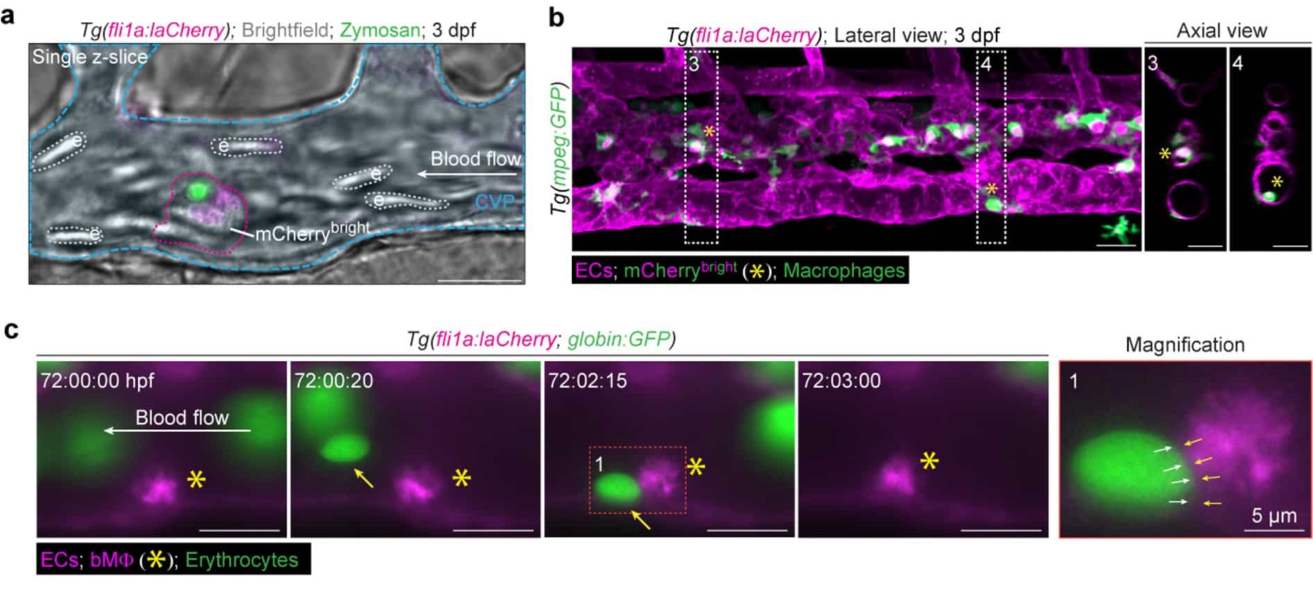
One of the most important lessons from this project is that imaging observation preceded molecular understanding. Transcriptomic and proteomic profiling later established that bMFs expressed both canonical macrophage and endothelial markers, confirming a hybrid endothelial-macrophage identity. Most strikingly, unlike virtually all known tissue-resident macrophages, bMFs did not express the macrophage receptor Csf1r, suggesting that both their production and function are independent of a signaling pathway widely considered indispensable for macrophage biology. Consistent with this prediction, csf1r mutant zebrafish had normal numbers of fully functional bMFs. Because CSF1R is commonly used to identify macrophages, its absence on bMFs may explain why this population has been overlooked by previous transcriptomics and conventional marker-based approaches.
Our curiosity then shifted to the origin of these cells. Once again, the answer came from our high spatiotemporal resolution real time imaging. By continuously imaging embryos from the onset of circulation, we observed individual endothelial cells change shape, detach from the vessel wall and enter the bloodstream, where they immediately adopted the characteristic behavior of bMFs. Remarkably, this transition occurred without any apparent progenitor intermediates. Instead, endothelial cells directly converted into bMFs, therefore we named this previously undescribed process endothelial-to-macrophage transition (EMacT).
Ironically, the project also answered a different question. We had long been puzzled by the poor efficiency of HSPC transplantation in zebrafish embryos. Fluorescent donor HSPCs injected into the circulation are expected to survive long enough to home to the caudal hematopoietic tissue, where they should expand before colonizing definitive hematopoietic organs such as the thymus and kidney (the zebrafish equivalent of mammalian bone marrow). Instead, most cells disappeared from the circulation within a few hours. Only after discovering bMΦs did we finally understand why. Live imaging showed us that donor HSPCs were intercepted almost immediately after entering the bloodstream, inspected, and frequently eliminated by bMΦs.
For us, this project became a reminder of something that is easy to forget in the era of large-scale omics datasets: sometimes seeing truly does come before knowing. Every major discovery in this study began with an unexpected observation under the microscope. Live imaging in zebrafish embryos allowed us to discover a cell population that no molecular snapshot could have predicted. Every major step in this project, from identifying these cells to uncovering their functions, to revealing their developmental origin, began with simply watching biology unfold one cell at a time. The power of imaging is illustrated by the 17 annotated supplementary videos accompanying our publication. Together, they allow readers to experience the discovery much as we did: by watching individual cells interact, move, and surprise us in real time. We are now exploring the broader roles of bMΦs in homeostasis and disease and investigating whether equivalent bMΦs exist in mammals, including humans. If this project taught us one lesson, it is that sometimes the most important discoveries are already there, we simply have to keep watching.
Check out the full research article for more details.
Get involved
Create an account or log in to post your story on FocalPlane.
More posts like this

Filter by
- NewsApply
- DiscussionsApply
- How toApply
- ToolsApply
- Case studiesApply
- InterviewsApply
- JobsApply
- EducationApply
- Blog seriesApply
- Volume EMApply
- Latin American Micro..scopistsApply
- Bio-image Analysis w..ith NapariApply
- Imaging with…Apply
- Towards Global Acces..sApply
- Latin America Bioima..gingApply
- From Zero to Qupath ..HeroApply
- Asian Microscopists ..and Cell BiologistsApply
- AIC at HHMI JaneliaApply
- Deep Learning for Bi..o-image analysisApply
- GloBIAS – updates fr..om the communityApply
- WAMBIAN: West Africa.. in FocusApply
- Highlights from Euro..-BioImagingApply
- LSFM seriesApply
- DIY MicroscopyApply
- View all