

Imaging spotlight: the role of intestinal muscles in remodelling the intestine
Posted by FocalPlane, on 28 October 2026
In this Imaging spotlight, Alessandro Mineo and Irene Miguel-Aliaga highlight the key findings from their research looking at the role of intestinal muscles in remodelling the intestine. They talk about developing the protocols and pipelines that allowed them to image and segment whole intestines. In the future, they’d like to be able to combine whole-organ imaging with cellular resolution in an intact, live animal.
What are the key results from your paper?
Until recently we thought that the intestinal muscles were just driving contractions and propelling the food along the gut, our findings showed that they can do much more: they can resize the entire intestine. In flies, males and females have different intestinal sizes and we found that the intrinsic sexual identity of their gut muscles contributes to these differences. This becomes particularly important for females during reproduction, as their intestinal muscles remodel to enlarge the intestine and sustain fecundity. Interestingly, this reproductive remodelling is not unique to flies. In mice, intestinal muscles also grow during pregnancy and even further during lactation. Intestinal plasticity has traditionally been attributed to the intestinal epithelium and its ability to self-renew, regenerate and resize in response to changing conditions. Our work adds the surrounding muscles to this picture, revealing them as active players in controlling intestinal size and adaptation. Given that many of our internal organs are surrounded by muscles, this raises the possibility that similar mechanisms could contribute to the remodelling of other organs during major physiological transitions, such as pregnancy or ageing.
Which imaging and image analysis techniques have you used in your research?
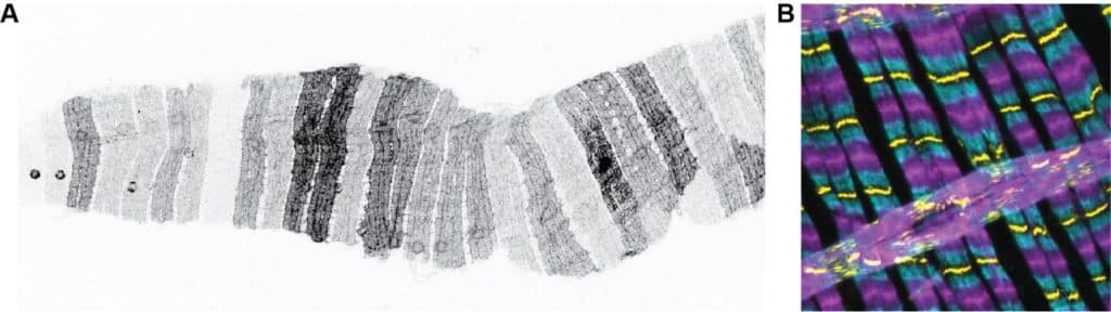
The projects required quite a broad range of imaging approaches, partly because these muscles were still poorly characterised. We used confocal microscopy with fluorescently labelled components of the contractile machinery, and we also did serial block-face electron microscopy on fixed samples, in collaboration with the Frank Schnorrer’s group in France, to characterise the muscle structure and understand how it changes between the sexes and during reproduction. We also wanted to understand how these muscles actually contract in vivo, and whether their contractile activity changes with sex or reproductive state. To find out, we used a method for imaging fly organs through the insect cuticle, which let us monitor muscle peristalsis in live animals.. We labelled the muscle nuclei with a strong fluorescent marker (Stinger::GFP) that could be detected through the insect cuticle, allowing us to follow the muscle peristaltic waves of contraction in intact animals.
Our imaging approaches also required developing a range of image analysis approaches. We had to quantify the organisation of the contractile machinery across many conditions and genotypes, so we developed an imaging pipeline to systematically and unbiasedly quantify muscle structure. Once we found that the muscles could influence the size of the entire intestine, we also wanted to understand how this affected the surrounding tissues. We therefore imaged whole intestines and used Cellpose for 3D segmentation and volumetric analysis. Finally, we were interested in how the sexual and reproductive state of the muscles affects the vascular-like tracheal network surrounding the gut, so we generated 3D reconstructions of the tracheal filaments using Imaris Filament Tracer.
Were there any technical challenges that you had to overcome in this research?
One of the most challenging aspects about this project was that we were working with a muscle system that had not been very well characterised, so we often had to develop or adapt approaches ourselves. A major challenge was simply being able to visualise the muscle contractile machinery. Many of the standard imaging approaches and markers didn’t work well, including antibodies commonly used to label contractile proteins in both flies and mammals. We therefore had to optimise markers and imaging conditions, as well as developing ways to image and segment whole intestines, which change dramatically in size and shape during reproduction.
Imaging muscle contraction in vivo was probably one of the most exciting technical challenges. The muscles contract very rapidly and are located inside the animal, so they are difficult to visualise through the cuticle. We found that labelling the muscle nuclei with a very strong fluorescent marker gave us a much cleaner signal. This allowed us to track the nuclei as the muscles contracted and, importantly, to quantify the frequency of peristaltic contractions in intact animals.
Are there any advances in imaging or image analysis that would help your research going forward?
One advance that would be particularly valuable for my research would be the ability to image gut muscle contractions across larger regions of the intestine, ideally the entire intestine, while maintaining high spatial and temporal resolution in live animals. At the moment, we can image muscle contractions in intact animals through the cuticle, but the resolution is not sufficient to resolve the cellular organisation of the muscles in detail. Being able to combine whole-organ imaging with cellular resolution in an intact, live animal would give us the unique opportunity to study the muscles in their natural environment, without dissecting or perturbing the intestinal physiology.
Read the full paper here: https://www.cell.com/cell/fulltext/S0092-8674(26)00822-6