Microscopy preprints – new tools and techniques in imaging
Posted by FocalPlane, on 7 February 2025
Here is a curated selection of preprints posted recently on new tools and techniques in imaging. Let us know if we are missing any recent preprints that are on your reading list!
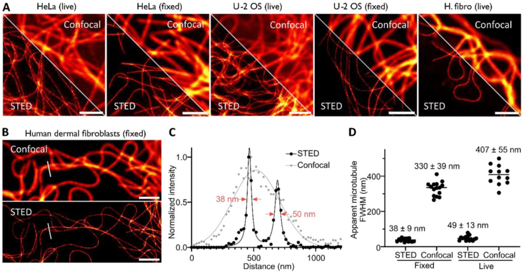
Biocompatible sulfonium-based covalent probes for endogenous tubulin fluorescence nanoscopy in live and fixed cells
Marie Auvray, Tanja Koenen, Olexandr Dybkov, Henning Urlaub, Gražvydas Lukinavičius
Optimizing multifunctional fluorescent ligands for intracellular labeling
Pratik Kumar, Jason D. Vevea, Ariana N. Tkachuk, Kirby Campbell, Emma T. Watson, Anthony X. Ayala, Jonathan B. Grimm, Edwin R. Chapman, David J. Solecki, Luke D. Lavis
Feature-Driven Whole-Tissue Imaging with Subcellular Resolution
Jinlong Lin, Zach Marin, Xiaoding Wang, Hazel M. Borges, Pierre-Emmanuel Y. N’Guetta, Xuemei Luo, Baylee A. Porter, Yuanyuan Xue, Md Torikul Islam, Tai Ngo, Arin B. Aurora, Hu Zhao, Suzanne D. Conzen, Sean J. Morrison, Shuang Liang, Zhenyu Zhong, Lori L. O’Brien, Kevin M. Dean
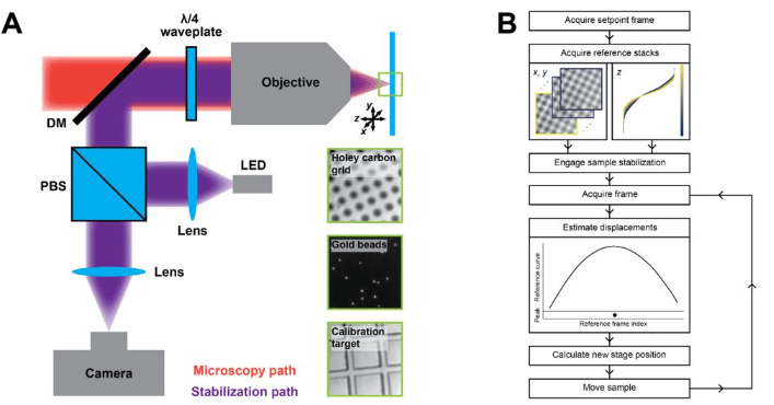
Image-based 3D active sample stabilization on the nanometer scale for optical microscopy
Jakob Vorlaufer, Nikolai Semenov, Caroline Kreuzinger, Manjunath G. Javoor, Bettina Zens, Nathalie Agudelo Dueñas, Mojtaba R. Tavakoli, Marek Šuplata, Wiebke Jahr, Julia Lyudchik, Andreas Wartak, Florian Schur, Johann G. Danzl
CALIPERS: Cell cycle-aware live imaging for phenotyping experiments and regeneration studies
Moises Di Sante, Melissa Pezzotti, Julius Zimmermann, Alessandro Enrico, Joran Deschamps, Elisa Balmas, Silvia Becca, Alessandro Reali, Alessandro Bertero, Florian Jug, Francesco S. Pasqualini
Single Objective Light Sheet Microscopy allows high resolution in vivo brain imaging of Drosophila
Francisco J. Tassara, Mariano Barella, Lourdes Simó, M. Mailén Folgueira Serrao, Micaela Rodríguez-Caron, Juan Ignacio Ispizua, Mark H. Ellisman, Horacio O. de la Iglesia, M. Fernanda Ceriani, Julián Gargiulo
Open-Source 3D Active Sample Stabilization for Fluorescence Microscopy
Sanket Patil, Giuseppe Vicidomini, Eli Slenders
An expanded palette of bright and photostable organellar Ca2+ sensors
Agathe Moret, Helen Farrants, Ruolin Fan, Kelsey Zingg, Christine E. Gee, Thomas G. Oertner, Vidhya Rangaraju, Eric R. Schreiter, Jaime de Juan-Sanz
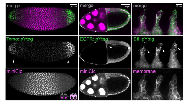
In vivo measurements of receptor tyrosine kinase activity reveal feedback regulation of a developmental gradient
Emily K. Ho, Rebecca P. Kim-Yip, Alison G. Simpkins, Payam E. Farahani, Harrison R. Oatman, Eszter Posfai, Stanislav Y. Shvartsman, Jared E. Toettcher
A highly photostable monomeric red fluorescent protein
Pingyong Xu, Ya Ding, Wenting He, Kunhao Wang, Fudong Xue, Shiqun Zhao, Leiting Pan, Liangyi Chen, Lin Yuan
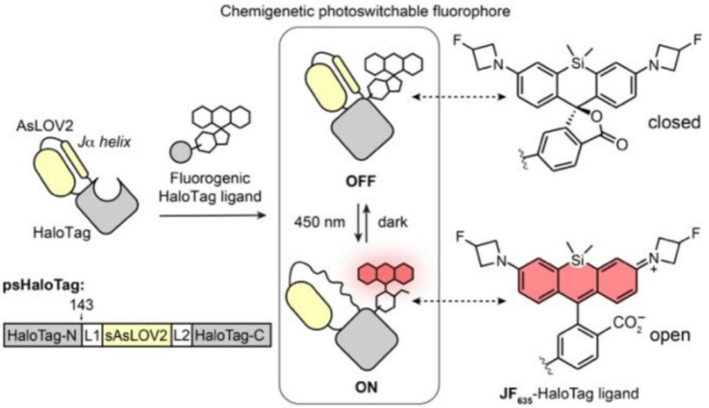
A photoswitchable HaloTag for spatiotemporal control of fluorescence in living cells
Franziska Walterspiel, Begoña Ugarte-Uribe, Jonas Weidenhausen, Anna Dimitriadi, Arif Ul Maula Khan, Christoph W. Müller, Claire Deo
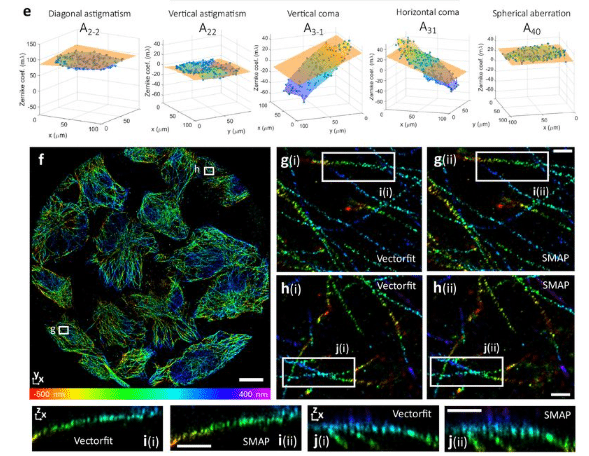
Calibration-free estimation of field dependent aberrations for single molecule localization microscopy across large fields of view
Isabel Droste, Erik Schuitema, Sajjad Khan, Stijn Heldens, Ben van Werkhoven, Keith A. Lidke, Sjoerd Stallinga, Bernd Rieger
Histology-Guided Single-Cell Mass Spectrometry Imaging using Integrated Bright-field and Fluorescence Microscopy
Alexander Potthoff, Marcel Niehaus, Sebastian Bessler, Jan Schwenzfeier, Emily Hoffmann, Oliver Soehnlein, Jens Höhndorf, Klaus Dreisewerd, Jens Soltwisch
Semi-automated navigation for efficient targeting of electron tomography to regions of interest in volume correlative light and electron microscopy
Kohki Konishi, Guilherme Neves, Matthew Russell, Masafumi Mimura, Juan Burrone, Roland Fleck
Fast Photostable Expansion Microscopy Using QDots and Deconvolution
Loku Gunawardhana, Wilna Moree, Jiaming Guo, Camille Artur, Tasha Womack, Jason L. Eriksen, David Mayerich
The dark side of fluorescent protein tagging – the impact of protein tags on biomolecular condensation
Edoardo Fatti, Sarah Khawaja, Karsten Weis
Single Objective Light Sheet Microscopy allows high resolution in vivo brain imaging of Drosophila
Francisco J. Tassara, Mariano Barella, Lourdes Simó, M. Mailén Folgueira Serrao, Micaela Rodríguez-Caron, Juan Ignacio Ispizua, Mark H. Ellisman, Horacio O. de la Iglesia, M. Fernanda Ceriani, Julián Gargiulo
Wavefront estimation through structured detection in laser scanning microscopy
Francesco Fersini, Alessandro Zunino, Pietro Morerio, Francesca Baldini, Martin J. Booth, Alessio Del Bue, Giuseppe Vicidomini
Increasing the acquisition speed in oblique plane microscopy via Aliasing
Conor Mcfadden, James Manton, Reto Fiolka
SurFlex Microscopy: Measuring Flexibility of Surface-Tethered Biomolecules
Aymeric Chorlay, Siddhansh Agarwal, Lena Blackmon, Daniel A. Fletcher
Outcome-Driven Microscopy: Closed-Loop Optogenetic Control of Cell Biology
Josiah B. Passmore, Alfredo Rates, Jakob Schröder, Menno T. P. van Laarhoven, Vincent J. W. Hellebrekers, Henrik G. van Hoef, Antonius J. M. Geurts, Wendy van Straaten, Wilco Nijenhuis, Florian Berger, Carlas S. Smith, Ihor Smal, Lukas C. Kapitein
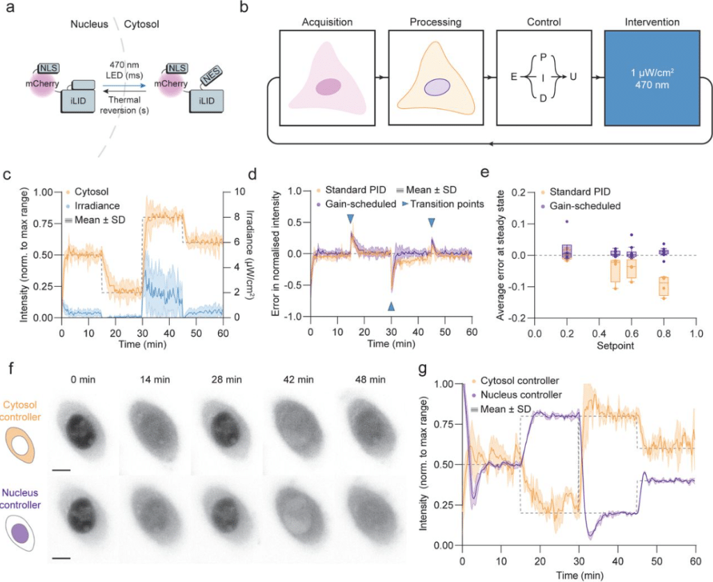
Figure extracted from Passmore, et al. The image is made available under a CC-BY-NC 4.0 International license.
FLIPs: Novel Genetically Encoded Biosensors for Functional Imaging of Cell Signaling by Polarization Microscopy
Paul Miclea, Vendula Nagy-Marková, Robin Van den Eynde, Wim Vandenberg, Alina Sakhi, Alexey Bondar, Jitka Myšková, Peter Dedecker, Josef Lazar
Time-resolved fluorescent proteins towards fluorescence microscopy in the temporal and spectral domains
Zizhu Tan, Chia-Heng Hsiung, Jiahui Feng, Yangye Zhang, Junlin Chen, Ke Sun, Peilong Lu, Jianyang Zang, Wenxing Yang, Ya Gao, Jiabin Yin, Tong Zhu, Yuxuan Ye, Yihan Wan, Xin Zhang
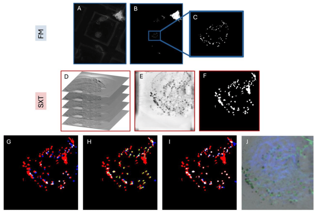
Demonstrating Soft X-Ray Tomography in the lab for correlative cryogenic biological imaging using X-rays and light microscopy
Stephen O’Connor, David Rogers, Maryna Kobylynska, James Geraets, Katja Thaysen, Jacob Marcus Egebjerg, Madeleen C. Brink, Louisa Herbsleb, Michaela Salakova, Leon Fuchs, Frauke Alves, Claus Feldmann, Axel Ekman, Paul Sheridan, William Fyans, Tony McEnroe, Fergal O’Reily, Kenneth Fahy, Roland A. Fleck, Daniel Wüstner, Jeremy C. Simpson, Andreas Walter, Sergey Kapishnikov
Polarization light-sheet microscopy and tomography (PLµTo) for flow-based imaging of 3D microcarrier mesenchymal stem cell culture
Oscar R. Benavides, Berkley P. White, Roland Kaunas, Carl A. Gregory, Alex J. Walsh
Adaptive-learning physics-aware light-field microscopy enables day-long and millisecond-scale super-resolution imaging of 3D subcellular dynamics
Lanxin Zhu, Jiahao Sun, Chengqiang Yi, Meng Zhang, Yihang Huang, Sicen Wu, Mian He, Liting Chen, Yicheng Zhang, Chunhong Zheng, Hao Chen, Yuhui Zhang, Dongyu Li, Peng Fei
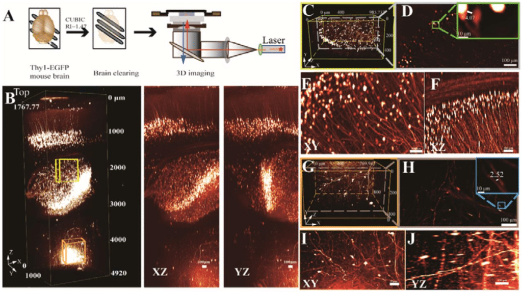
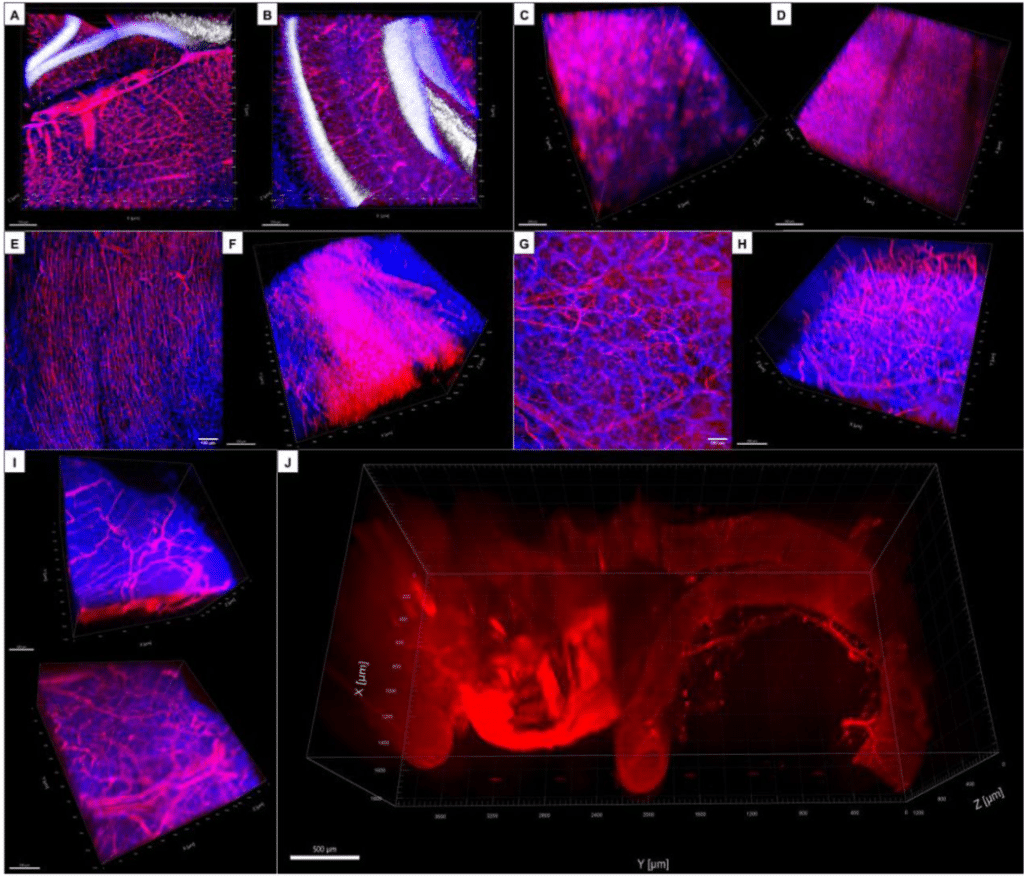
A novel method (RIM-Deep) for enhancing imaging depth and resolution stability of deep cleared tissue in inverted confocal microscopy
Yisi Liu, Pu Wang, Junjie Zou, Hongwei Zhou
Novel Laser Technology Enables 10x Faster SRS Imaging and Rapid Tuning in Biological Samples
Lenny Reinkensmeier, René Siegmund, Mark Bates, Gero Stibenz, Peter Trabs, Stefan Popien, Ingo Rimke, Sandro Heuke, Alexander Egner
Single-shot label-free nanoscopy for quantitative organelle visualization on standard commercial microscopes
Yugo Inutsuka, Koki Yamamoto, Masafumi Kuroda, Yasushi Okada
White and Clearing: New Optical Tissue Clearing Method
Vicente Llorente, Elena Fernández-Cortés, Daniel Sanderson, Julia Salazar, Jorge Ripoll, Manuel Desco, María Victoria Gómez-Gaviro
A fully 3D-printed optical microscope for low-cost histological imaging
Jay Christopher, Rebecca Craig, Rebecca E. McHugh, Andrew J. Roe, Ralf Bauer, Brian Patton, Gail McConnell, Liam M. Rooney
TWINKLE: An open-source two-photon microscope for teaching and research
Manuel Schottdorf, P. Dylan Rich, E. Mika Diamanti, Albert Lin, Sina Tafazoli, Edward H. Nieh, Stephan Y. Thiberge
I2SIM: Boosting High-Fidelity Isotropic Super-Resolution with Image Interference and Spatial-Spectral Optimization
Enxing He, Yile Sun, Hongfei Zhu, Xinxun Yang, Lu Yin, Yubing Han, Cuifang Kuang, Xu Liu
Single-Photon Single-Particle Tracking
Lance W.Q. Xu, Nathan Ronceray, Marianna Fanouria Mitsioni, Aleksandra Radenovic, Steve Pressé
Directionality of two-photon absorption in representative fluorescent proteins
Josef Lazar, Štěpán Timr, Olga Rybakova, Jiří Brynda, Adam Thuen, Mikhail Drobizhev, Jitka Myšková
3D imaging with superior resolution using Atacama Clear
Lauretta A. Lacko, Tansol Choi, Neranjan de Silva, Ying Liu, Edgar A. Jamies, Todd Evans, Romulo Hurtado
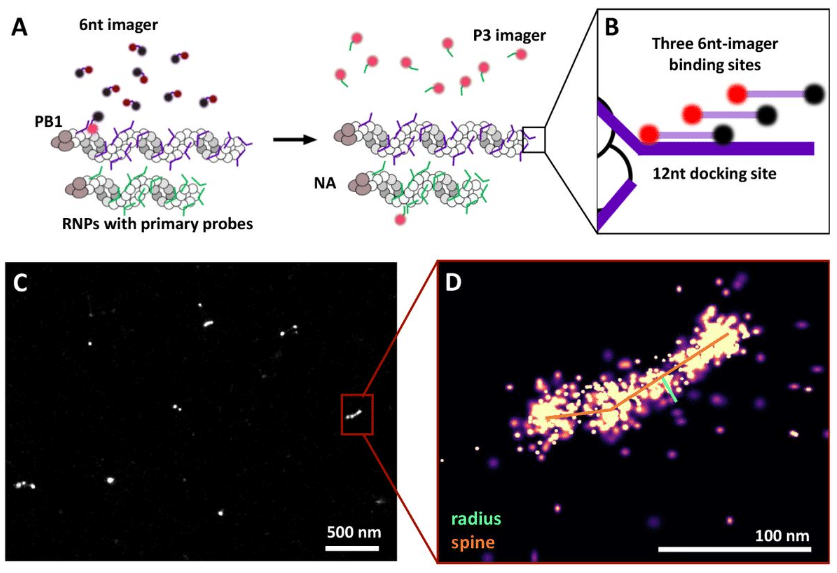
Tunable fluorogenic DNA probes drive fast and high-resolution single-molecule fluorescence imaging
Mirjam Kümmerlin, Qing Zhao, Jagadish Hazra, Christof Hepp, Alison Farrar, Piers Turner, Achillefs N. Kapanidis
All-Optical Strategies to Minimize Photo-Bleaching in Reversibly Switchable Fluorescent Proteins
Guillem Marín-Aguilera, Francesca Pennacchietti, Andrea Volpato, Alessia Papalini, Abhilash Kulkarni, Niusha Bagheri, Guillaume Minet, Jerker Widengren, Ilaria Testa
A Palette of Bridged Bicycle-Strengthened Fluorophores
Junwei Zhang, Kecheng Zhang, Kui Wang, Bo Wang, Siyan Zhu, Hongping Qian, Yumiao Ma, Mengling Zhang, Tianyan Liu, Peng Chen, Yuan Shen, Yunzhe Fu, Shilin Fang, Xinxin Zhang, Peng Zou, Wulan Deng, Yu Mu, Zhixing Chen
Ether Rhodamines with Enhanced Hydrophilicity, Fluorogenicity, and Brightness for Super-Resolution Imaging
Xiangning Fang, Qinglong Qiao, Zhifeng Li, Hao-Kai Li, Jie Chen, Ning Xu, Kai An, Wenchao Jiang, Yi Tao, Pengjun Bao, Yinchan Zhang, Zhimin Wu, Xiaogang Liu, Zhaochao Xu
SiR-XActin: A fluorescent probe for imaging actin dynamics in live cells
Veselin Nasufovic, Julian Kompa, Halli L. Lindamood, Merle Blümke, Birgit Koch, Victoria Le-vario-Diaz, Katharina Weber, Marlene Maager, Elisabetta Ada Cavalcanti-Adam, Eric A. Vitriol, Hans-Dieter Arndt, Kai Johnsson
 (No Ratings Yet)
Get involved
Create an account or log in to post your story on FocalPlane.
More posts like this

Filter by
- NewsApply
- DiscussionsApply
- How toApply
- ToolsApply
- Case studiesApply
- InterviewsApply
- JobsApply
- EducationApply
- Blog seriesApply
- Volume EMApply
- Latin American Micro..scopistsApply
- Bio-image Analysis w..ith NapariApply
- Imaging with…Apply
- Towards Global Acces..sApply
- Latin America Bioima..gingApply
- From Zero to Qupath ..HeroApply
- Asian Microscopists ..and Cell BiologistsApply
- AIC at HHMI JaneliaApply
- Deep Learning for Bi..o-image analysisApply
- GloBIAS – updates fr..om the communityApply
- Highlights from Euro..-BioImagingApply
- LSFM seriesApply
- DIY MicroscopyApply
- View all