Featured image with Brittany Carr
Posted by FocalPlane, on 24 April 2026
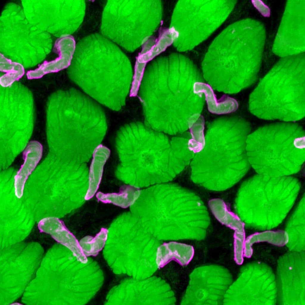
Our featured image, acquired by Brittany Carr, shows the rod and cone photoreceptor outer segments from a tadpole retina.
Photoreceptor outer segments are responsible for capturing photons of light and transducing them into chemical signals. Frog rods are large, so this image also shows the incisures, which are invaginations into the outer segment that gives it a flower-like appearance. This image was prepared by immunofluorescence using a lectin (wheat germ agglutinin, green) and a cone opsin antibody (magenta). It was imaged using pseudo-super-resolution light microscopy (Airyscan). They are shown here at about 1200x magnification.
Discover more about Brittany’s research.
Research career so far: During my PhD research, I was interested in pharmacological control of myopia, and investigating off-target drug effects of muscarinic antagonists in the chicken eye. I then switched to inherited retinal degeneration for my postdoctoral studies, where I learned to perform genetic modification with CRISPR and to use frogs as a model organism. I studied the effects of loss of two genes: PROM1 and CDHR1 on photoreceptor outer segment morphogenesis and retinal degeneration.
Current research: I started my independent research career in 2022. I am still interested in the developmental and degenerative effects of PROM1 variants, and in using frogs to develop other models of inherited and age-related blindness. We are developing a few other interesting projects in the lab involving other cell types in the retina, including microglia and Müller glia.
Favourite imaging technique: From the first time I took a simple micrograph of a dopaminergic amacrine cell, I was hooked. I love confocal and super-resolution light microscopy. It is a simple technique and you can create such beautiful images of nature. It’s a very fulfilling way for me to combine my love of science and art.
What are you most excited about in microscopy? I think that, to me, confocal and super-resolution light microscopy will always be king for making beautiful images, but spatial transcriptomics (MERFISH) is an exciting technique. The ability to look at RNA expression across entire tissues, instead of single cells, is just incredible to me, and I think that it will lead to interesting insights into how cellular and genetic networks work together to support entire tissues or organisms.
 (No Ratings Yet)