Featured image with Joan Roncero Carol
Posted by FocalPlane, on 3 July 2026
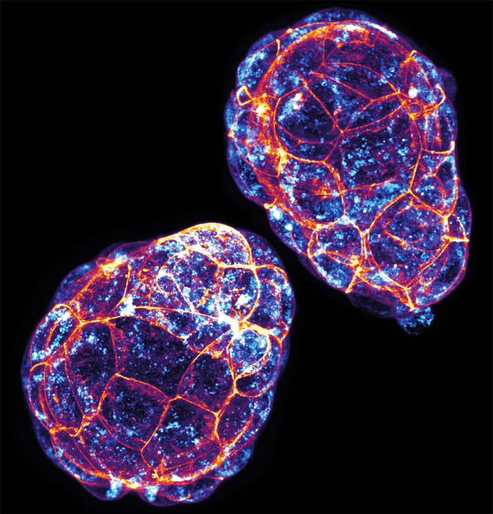
Our featured image, acquired by Joan Roncero Carol, shows two mouse preimplantation blastocysts at E4.5 with immunofluorescence staining against LAMP1 to visualize lysosomal distribution at this developmental stage, together with phalloidin staining to clearly outline cell membranes. The image was acquired using a Zeiss LSM 980 confocal microscope with the Airyscan module and subsequently processed using ImageJ.
Discover more about Joan’s research.
Research career so far: My research has focused on early embryonic development and embryo–environment interactions. During my PhD in Dr. Hoijman’s lab at IDIBELL and IBMB-CSIC in Barcelona, I have studied how early embryos interact with bacteria using zebrafish, mouse and human donated embryos combined with advanced imaging approaches.
Current research: I am currently studying whether preimplantation embryos have innate immune-like capacities to respond to potential infections. In particular, I am investigating the mechanisms by which embryos detect, internalize, and process external agents such as bacteria, with a focus on the cellular and molecular pathways involved.
Favourite imaging technique/microscope: I particularly enjoy live imaging, as it allows me to visualize cellular and tissue dynamics in real time in living embryos.
What are you most excited about in microscopy? I really like confocal microscopy, and I am excited to further explore super-resolution techniques to visualize cellular dynamics in much greater detail. Being able to visualize the protrusions that embryos extend to capture bacteria in much finer detail would be amazing!
 (1 votes, average: 1.00 out of 1)