

Featured image with Ádám Soós
Posted by FocalPlane, on 25 September 2026
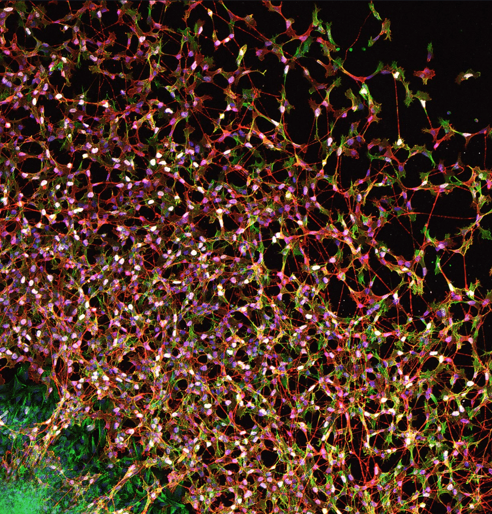
Our featured image, acquired by Ádám Soós is titled “Guided by GDNF: enteric neural crest cells weaving the future gut nervous system.” It shows enteric neural crest-derived cells streaming out from an embryonic day 6 chicken midgut explant, forming an interconnected migratory network on a fibronectin-coated surface. The culture was exposed to 20 ng/ml GDNF, a chemoattractant that guides the outgrowth of these cells as they colonize their environment and begin to assemble a cellular architecture similar to that of the enteric nervous system.
The image captures both movement and identity. TUJ1 staining in red highlights neuronal differentiation and extended neurites, while HNK1 in green marks migratory neural crest-derived cells. EdU Click-iT labelling appears in white, revealing proliferating nuclei within the expanding population, and DAPI in blue counterstains all nuclei. Together, the channels create a dense constellation of migrating, dividing and differentiating cells, evoking the emergence of the gut’s intrinsic nervous system as a living, self-organizing network in vitro.
Firstly, the midgut was isolated from the chicken embryo. The midgut was explanted on a fibronectin-coated Petri dish, and GDNF was added to DMEM media. Secondly, EdU labelling was made in the last 3 hours. After that, the sample was 3% PFA-fixed after 24 hours. After washing steps, it was immunolabelled for TUJ1, HNK1, EdU and DAPI. The image was acquired by laser-scanning confocal microscopy using a Zeiss LSM 780 microscope with a 20x objective. The image was processed for channel visualization, contrast adjustment and final presentation.
Read more about Ádám’s research.
Research career so far: My research career has been shaped by developmental biology, embryology and microscopy. I completed my BSc in Biology at Babeș-Bolyai University in Cluj-Napoca, where I worked on the growth dynamics and biomass production of the green microalga Tetradesmus obliquus. I then completed my MSc in Biology at Eötvös Loránd University, specializing in molecular, immune and microbiology. During this period, I became interested in embryonic organ development and worked on the cellular and molecular characterization of the bursa of Fabricius.
Since 2021, I have been a PhD student at Semmelweis University in the Doctoral School of Molecular Medicine, working in the Stem Cell and Experimental Embryology Laboratory under the supervision of Prof. Dr. Nándor Nagy. My work has focused on embryonic development, lymphoid organogenesis, extracellular matrix biology and, more recently, enteric nervous system development. Along the way, I have gained experience in sample preparation techniques, embryonic manipulation, organoid cultures, immunohistochemistry, in vitro, ex vivo and in ovo culture techniques, confocal- and superresolution microscopy, electron microscopy, antibody production, virus transfection and other techniques.
I also completed a research internship for 6 months at the Centre for Brain Research (CBR), Medical University of Vienna, in the Adameyko Laboratory, where I worked on human embryonic neural crest cells as sources of sympathoadrenal tumours using iPSC culture techniques, organoid techniques, virus transfection and bioinformatics.
Current research: My current research focuses on the embryonic development of the enteric nervous system, particularly how the microenvironment guides enteric neural crest-derived cells during migration, proliferation and differentiation. I use avian embryonic models, in vitro explant cultures, neurosphere-based approaches, transplantation experiments and advanced imaging to study how growth factors and extracellular matrix components influence the colonization of the developing gut.
A central aim of my work is to understand how enteric neural stem cells interact with their surrounding environment and how this knowledge could support future regenerative approaches for enteric nervous system disorders. My recent work includes studying how cecal growth factors promote enteric neurosphere formation and hindgut colonization in the avian model.
Favourite imaging technique/microscope: My favourite imaging techniques are live-cell imaging with fluorescent markers and transmission electron microscopy. Live-cell imaging is fascinating because it allows us to follow dynamic biological processes as they happen, especially cell migration, interaction and tissue organization. Transmission electron microscopy is equally exciting because it reveals another dimension of biology, showing ultrastructural details that very few people get to see directly.
What are you most excited about in microscopy? I am very excited about light-sheet microscopy and expansion microscopy. With these approaches, we can observe cells within their native environment at high resolution and across larger tissue volumes. I think this combination of context and detail will be extremely powerful for developmental biology, because it allows us to ask biologically meaningful questions not only about individual cells, but also about how cells communicate, organize and build tissues.