Microscopy preprints – applications in biology
Posted by FocalPlane, on 21 February 2025
Here is a curated selection of preprints published recently. In this post, we share preprints that use microscopy tools to answer questions in biology.
High-Resolution Structures of Tobacco Mosaic Virus Disks from Cryo-Electron Microscopy
Ismael Abu-Baker, Artur P. Biela, Sachin N. Shah, Jonathan G. Heddle, George P. Lomonossoff, Amy Szuchmacher Blum
Memory engram synapse 3D molecular architecture visualized by cryoCLEM-guided cryoET
Charlie Lovatt, Thomas J. O’Sullivan, Clara Ortega-de San Luis, Tomás J. Ryan, René A. W. Frank
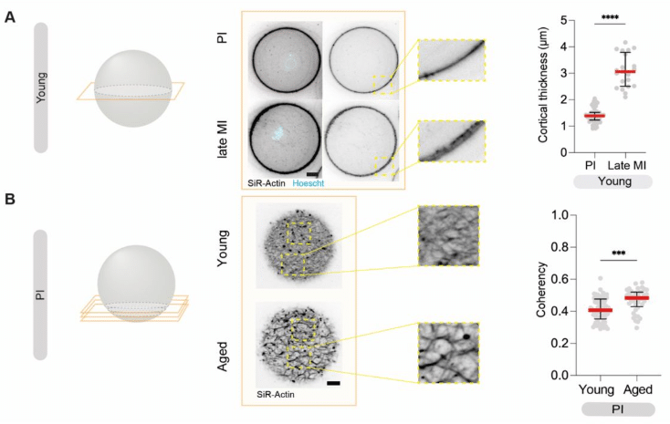
Atomic Force Microscopy reveals differences in mechanical properties linked to cortical structure in mouse and human oocytes
Rose Bulteau, Lucie Barbier, Guillaume Lamour, Yassir Lemseffer, Marie-Hélène Verlhac, Nicolas Tessandier, Elsa Labrune, Martin Lenz, Marie-Emilie Terret, Clément Campillo
Fibroblast contractility drives network reorganization and epithelial proliferation in intestinal polyposis
Mei-Lan Li, Yuying Wang, Maria Figetakis, David Gonzalez, Jason Jin, Elizabeth S. McDonald, Nadia A. Ameen, Kaelyn Sumigray
Mitochondria transported by Kinesin 3 prevent localized calcium spiking to inhibit caspase-dependent specialized cell death
Rashna Sharmin, Aladin Elkhalil, Sara Pena, Pranya Gaddipati, Ginger Clark, Pavak K. Shah, Mark W. Pellegrino, Shai Shaham, Piya Ghose
Image-based screens identify regulators of endogenous Dvl2 biomolecular condensates
Antonia Schubert, Florian Heigwer, Christian Scheeder, Oksana Voloshanenko, Dominique Kranz, Franziska Ragaller, Nadine Winkler, Thilo Miersch, Barbara Schmitt, Melanie Kuhse, Daniel Gimenes, Diana Ordoñez-Rueda, Jennifer Schwarz, Frank Stein, Dirk Jäger, Ulrike Engel, Michael Boutros
Fluorescence lifetime imaging microscopy for metabolic analysis of LDHB inhibition in triple negative breast cancer
A. Galloway, B. Ter Hofstede, Alex J. Walsh
Timelapse and volumetric imaging of mitochondrial networking using NAD(P)H autofluorescence via 2-photon microscopy
Blanche ter Hofstede, Alex J. Walsh
Quantifying the organization and dynamics of M. smegmatis morphology from Long-Term Time-Lapse Atomic Force Microscopy
Clément Soubrier, Anotida Madzvamuse, Haig Alexander Eskandarian, Khanh Dao Duc
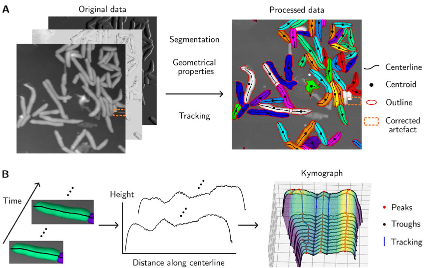
Figure extracted from Soubrier, et al. The image is made available under a CC-BY 4.0 International license.
Quantitative Spatial Analysis of Chromatin Biomolecular Condensates using Cryo-Electron Tomography
Huabin Zhou, Joshua Hutchings, Momoko Shiozaki, Xiaowei Zhao, Lynda K. Doolittle, Shixin Yang, Rui Yan, Nikki Jean, Margot Riggi, Zhiheng Yu, Elizabeth Villa, Michael K. Rosen
Cryogenic electron tomography and elemental analysis of mitochondrial granules in human retinal ganglion cells
Gong-Her Wu, Cathy Hou, Andrew Thron, Hirenkumar Rajendra Patel, Liam Spillane, Sanket Rajan Gupte, Serena Yeung-Levy, Sahil Gulati, Christopher Booth, Yaping Joyce Liao, Wah Chiu
Real-Time Analysis of Nanoscale Dynamics in Membrane Protein Insertion via Single-Molecule Imaging
C. Yang, D. Ma, S. Hu, M. Li, Y. Lu
Fluorescence Lifetime Imaging Microscopy (FLIM) visualizes internalization and biological impact of nanoplastics in live intestinal organoids
Irina A. Okkelman, Hang Zhou, Sergey M. Borisov, Angela C. Debruyne, Austin E. Y. T. Lefebvre, Marcelo Leomil Zoccoler, Linglong Chen, Bert Devriendt, Ruslan I. Dmitriev
Insect wings arose with a genetic circuit that extends the useful range of a BMP morphogen
Anqi Huang, Luca Cocconi, Ben Nicholls-Mindlin, Cyrille Alexandre, Guillaume Salbreux, Jean-Paul Vincent
Ångström-resolution imaging of cell-surface glycans
Luciano A. Masullo, Karim Almahayni, Isabelle Pachmayr, Monique Honsa, Larissa Heinze, Sarah Fritsche, Heinrich Grabmayr, Ralf Jungmann, Leonhard Möckl
Polarity reversal of stable microtubules during neuronal development
Malina K. Iwanski, Albert K. Serweta, H. Noor Verwei, Bronte C. Donders, Lukas C. Kapitein
Cryo-correlative light and electron tomography of dopaminergic axonal varicosities reveals non-synaptic modulation of cortico-striatal synapses
Paul Lapios, Robin Anger, Vincent Paget-Blanc, Esther Marza, Vladan Lučić, Rémi Fronzes, Etienne Herzog, David Perrais
Cell heterogeneity and fate bistability drive tissue patterning during intestinal regeneration
C. Schwayer, S. Barbiero, D. B. Brückner, C. Baader, N. A. Repina, O. E. Diaz, L. Challet Meylan, V. Kalck, S. Suppinger, Q. Yang, J. Schnabl, U. Kilik, J. G. Camp, B. Stockinger, M. Bühler, M. B. Stadler, E. Hannezo, P. Liberali
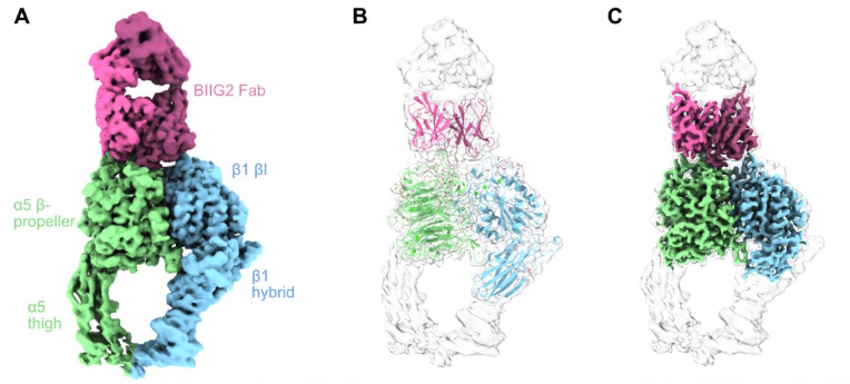
Structural and functional characterization of integrin α5-targeting antibodies for anti-angiogenic therapy
Adam Nguyen, Joel B. Heim, Gabriele Cordara, Matthew C. Chan, Hedda Johannesen, Cristine Charlesworth, Ming Li, Caleigh M. Azumaya, Benjamin Madden, Ute Krengel, Alexander Meves, Melody G. Campbell
Distinct filament morphology and membrane tethering features of the dual FtsZs in Odinarchaeota
Jayanti Kumari, Akhilesh Uthaman, Ananya Kundu, Anubhav Dhar, Vaibhav Sharma, Sucharita Bose, Soumyajit Dutta, Srijita Roy, Ramanujam Srinivasan, Samay Pande, Kutti R. Vinothkumar, Pananghat Gayathri, Saravanan Palani
Geometry-driven asymmetric cell divisions pattern cell cycles and zygotic genome activation in the zebrafish embryo
Nikhil Mishra, Yuting I. Li, Edouard Hannezo, Carl-Philipp Heisenberg
Lipid packing and local geometry influence septin curvature sensing
Brandy N. Curtis, Ellysa J. D. Vogt, Christopher Edelmaier, Amy S. Gladfelter
Spatiotemporal temperature control by holographic heating microscopy unveils cellular thermosensitive calcium signaling
Kotaro Oyama, Ayumi Ishii, Shuhei Matsumura, Tomoko Gowa Oyama, Mitsumasa Taguchi, Madoka Suzuki
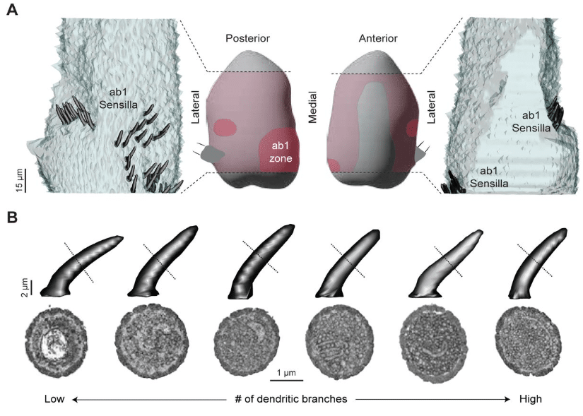
Population-level morphological analysis of paired CO2- and odor-sensing olfactory neurons in D. melanogaster via volume electron microscopy
Jonathan Choy, Shadi Charara, Kalyani Cauwenberghs, Quintyn McKaughan, Keun-Young Kim, Mark H. Ellisman, Chih-Ying Su
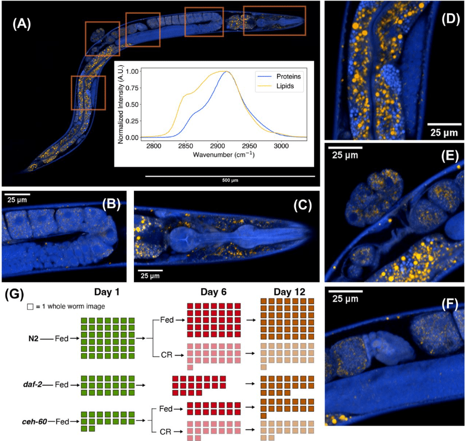
SRS microscopy identifies inhibition of vitellogenesis as a mediator of lifespan extension by caloric restriction in C. elegans
Bowen Yang, Bryce Manifold, Wuji Han, Catherin DeSousa, Wanyi Zhu, Aaron Streets, Denis V. Titov
Microscopy-Guided Spatial Proteomics Reveals Novel Proteins at the Mitochondria-Lipid Droplet Interface and Their Role in Lipid Metabolism
Yen-Ming Lin, Weng Man Chong, Chun-Kai Huang, Hsiao-Jen Chang, Chantal Hoi Yin Cheung, Jung-Chi Liao
Actomyosin and the Arp2/3 Complex Are Involved in the Internalization of Cellulose Synthase Complexes
Liyuan Xu, Weiwei Zhang, Lei Huang, Chunhua Zhang, Christopher J. Staiger

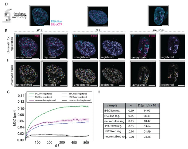
Super-resolution compatible DNA labeling technique reveals chromatin mobility and organization changes during differentiation
Maruthi K. Pabba, Miroslav Kuba, Tomáš Kraus, Kerem Celikay, Janis Meyer, Sunik Kumar Pradhan, Andreas Maiser, Hartmann Harz, Heinrich Leonhardt, Karl Rohr, Michal Hocek, M. Cristina Cardoso
Super-resolution microscopy of mitochondrial mRNAs
Stefan Stoldt, Frederike Maass, Michael Weber, Sven Dennerlein, Peter Ilgen, Jutta Gärtner, Aysenur Canfes, Sarah V. Schweighofer, Daniel C. Jans, Peter Rehling, Stefan Jakobs
Ultrastructure expansion microscopy of axonemal dynein in islet primary cilia
Xinhang Dong, Jeong Hun Jo, Jing Hughes
Mapping Alzheimer Disease Molecular Pathologies in Large-Scale Connectomics Data: A Publicly Accessible Correlative Microscopy Resource
Xiaomeng Han, Peter H. Li, Shuohong Wang, Tim Blakely, Sneha Aggarwal, Bhavika Gopalani, Morgan Sanchez, Richard Schalek, Yaron Meirovitch, Zudi Lin, Daniel Berger, Yuelong Wu, Fatima Aly, Sylvie Bay, Benoît Delatour, Pierre Lafaye, Hanspeter Pfister, Donglai Wei, Viren Jain, Hidde Ploegh, Jeff Lichtman
Scanning Electron Microscopy Study of Bacterial Growth in Mycelial Extracellular Matrices
Davin Browner, Andrew Adamatzky

GLP-1R associates with VAPB and SPHKAP at ERMCSs to regulate β-cell mitochondrial remodelling and function
Gregory Austin, Affiong I. Oqua, Liliane El Eid, Mingli Zhu, Yusman Manchanda, Priyanka Peres, Helena Coyle, Yelyzaveta Poliakova, Karim Bouzakri, Alex Montoya, Dominic J. Withers, Michele Solimena, Ben Jones, Steven J. Millership, Steffen Burgold, David C.A. Gaboriau, Endre Majorovits, Inga Prokopenko, Jonathon Nixon-Abell, Andreas Müller, Alejandra Tomas
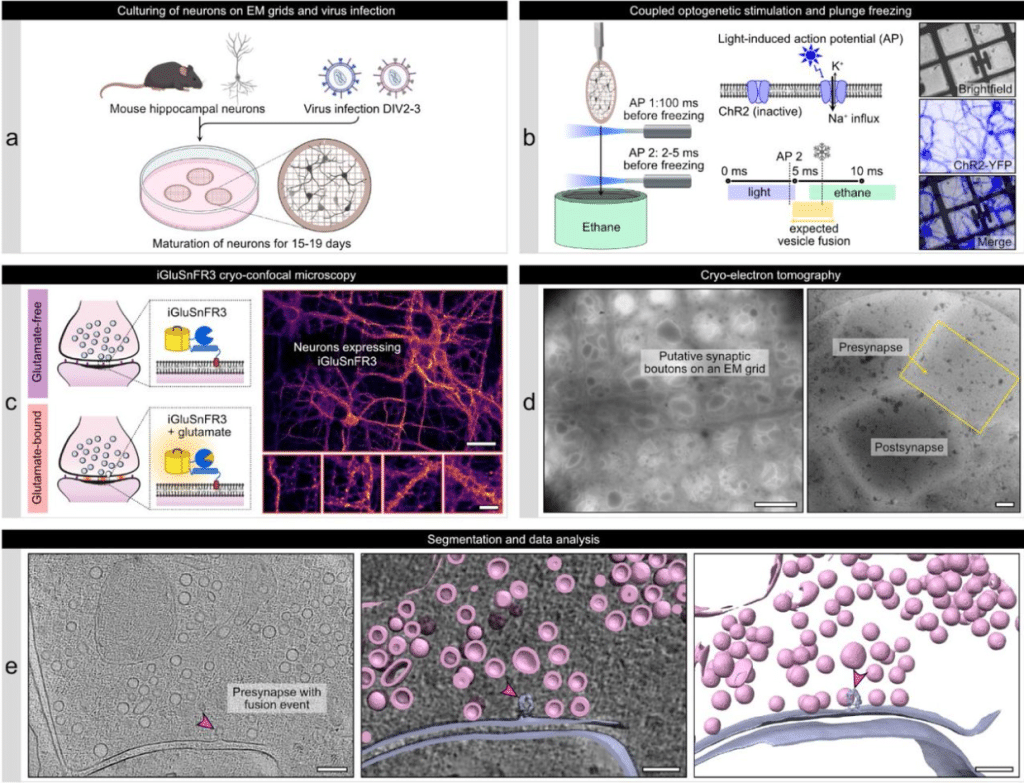
Dynamic nanoscale architecture of synaptic vesicle fusion in mouse hippocampal neurons
Jana Kroll, Uljana Kravčenko, Mohsen Sadeghi, Christoph A. Diebolder, Lia Ivanov, Małgorzata Lubas, Thiemo Sprink, Magdalena Schacherl, Mikhail Kudryashev, Christian Rosenmund
 (No Ratings Yet)
Get involved
Create an account or log in to post your story on FocalPlane.
More posts like this

Filter by
- NewsApply
- DiscussionsApply
- How toApply
- ToolsApply
- Case studiesApply
- InterviewsApply
- JobsApply
- EducationApply
- Blog seriesApply
- Towards Global Acces..sApply
- Latin America Bioima..gingApply
- From Zero to Qupath ..HeroApply
- Asian Microscopists ..and Cell BiologistsApply
- AIC at HHMI JaneliaApply
- Deep Learning for Bi..o-image analysisApply
- GloBIAS – updates fr..om the communityApply
- WAMBIAN: West Africa.. in FocusApply
- Volume EMApply
- Latin American Micro..scopistsApply
- Bio-image Analysis w..ith NapariApply
- Imaging with…Apply
- Highlights from Euro..-BioImagingApply
- LSFM seriesApply
- DIY MicroscopyApply
- View all