Featured image with Kaiden Power
Posted by FocalPlane, on 26 September 2025
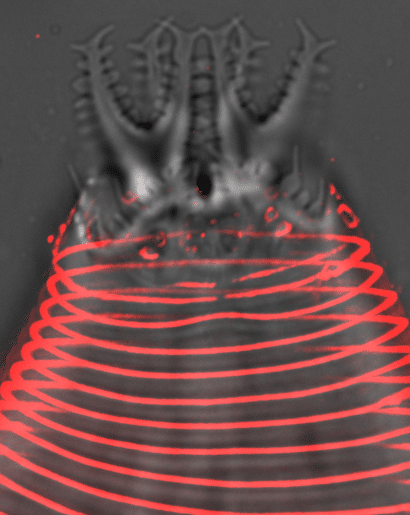
Our featured image, acquired by Kaiden Power, shows the head and probolae of an Acrobeles complexus nematode. The cuticle furrows (annuli) and cilia pores are stained with Concanavalin A (red). The sample was mounted and imaged live on a DeltaVision widefield microscope with deconvolution post-acquisition.
Discover more about Kaiden’s research.
Research career so far: I graduated from Ursinus College in 2015, where I studied polarity in the C. elegans early embryo. After graduation, I was a post-baccalaureate research fellow at the NIH studying how mitochondria change with age. For my thesis research at Rutgers University, I characterized novel roles for the protein NEKL-4/NEK10 in ciliary integrity and mitochondrial health, and obtained my PhD in January 2024. Currently, I am a postdoc at Boston Children’s Hospital’s Department of Genetics and Genomics.
Current research: My current research focuses on attachments between neurons and glia and how they affect neuron morphogenesis, as well as how these attachments are conserved or changed over evolution between different organisms.
Favourite imaging technique: My favourite imaging technique is confocal microscopy because of its versatility and high resolution, and my favourite microscope to use is the Zeiss LSM 880. My second favourite is the Zeiss Elyra SIM.
What are you most excited about in microscopy? I am most excited to learn new microscopy techniques during my postdoc and apply them to my research, specifically FRAP. In general I look forward to using new microscopes with improved resolution and deconvolution software suited to cell biological research.
 (No Ratings Yet)