Imaging spotlight: U-ExM atlas of microbial eukaryotes
Posted by FocalPlane, on 20 January 2026
In this paper highlight from Hiral Shah and colleagues, we learn about their ultrastructure expansion microscopy (U-ExM) atlas of over 200 cultured planktonic eukaryotes. This post includes technical advice for ExM users and highlights the open-access resource that they have generated for cytoskeleton or marine microorganism aficionados (and microscopy sciart lovers!)
What are the key results from your paper?
We capture the diversity of eukaryotic cellular organisation by generating an ultrastructure expansion microscopy (U-ExM)-based atlas of over 200 microbial eukaryotes. U-ExM expands cells ~4 times by embedding them in a swellable gel, offering a higher resolution on standard microscopes. Next, we use immuno-fluorescence for tubulin and centrin to visualize the diverse cytokeletal architectures. Combining this with protein pan-labelling with NHS-ester dyes that bind to all amine groups, and thus provide an overview of cellular organisation, we generate a map of cytoskeletal diversity across all the major eukaryotic groups. During the Roscoff and Bilbao stops of EMBL’s pan-European TREC expedition, we tapped into the diversity of the Roscoff and Basque microalgae culture collections, concentrating and fixing tens of cultures over days. Collectively, between the Dey, Dudin and Centriole Labs, we imaged hundreds of cells in 3D at high resolution, allowing us to observe new cytoskeletal structures as well as identify those only observed by EM previously. The fact that these antibodies worked across a wide range of species was a big step forward in developing this massive atlas. Along with Armando Rubio Ramos and Felix Mikus, we then annotated all images for cytoskeletal and subcellular features such as the architecture of cortical microtubule network, organisation of basal bodies, centrin rings and nucleolar-NE association. By imaging several species within lineages, as in the case of dinoflagellates, this resource lays the groundwork for future evolutionary studies and marine cell biology research across organism lifecycles and environmental fluctuations. Finally, we applied our pipeline to mixed environmental samples to discover amazing ultrastructural details within ciliates. All of these images are publicly available through the MoBIE plugin in Fiji. If you are interested in cytoskeleton, or weird marine species or just beautiful images, I totally recommend playing around with the dataset. With MoBIE, you can filter by species or groups of interest, zoom in and even download for further analysis. If you just want a quick look at the images, there is also a pdf available showcasing a curated set of species.
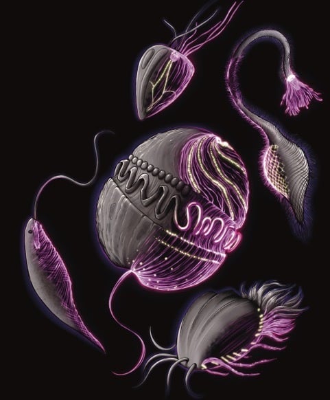
Illustration showing the outer surface of protists with a sci-fi style depiction of the internal cytoskeleton revealed in the U-ExM atlas.
Are there technical tips or tricks for getting started with U-ExM that you can share with us? Are you open to researchers contacting you for collaboration on the methodology?
The key step is making and handling the U-ExM gels. This can take some practice initially, but once you see the gorgeous images, it’s hard to stop using this technique and everyone finds their own little tools and ways to handle them. First of all, to get the gels right you want to pay attention to gel composition, quality of sodium-acrylate, cell density and immunostaining. You can find more such notes in Gambarotto et al, 2021 or in the more recent protocols.io. Shifting to smaller 6mm coverslips to make gels, for instance, allowed us to downscale reagents and made it easier to handle and store gels. Another big one was the use of plunge-freezing for cryo-fixation where possible (Laporte et al, 2022). In addition to preserving cell architecture better, it allows expansion of cells with tough cell walls, as in the case of diatoms (Flori, Mikus, Flaum, et al, 2025).
We are always open to helping researchers adopt the method and have been sharing the technique with visitors in the lab as well as through more structured courses in Europe and abroad. If you have a species and questions you are interested in trying ExM on and don’t know where to start, you can submit a request to ExAME or the GenExM facility at University of Geneva.
Are there any advances in ExM that you hope to see in the future?
Expansion microscopy relies on having good labelling strategies. So, we are always on the look out for new antibodies and dyes to label specific cellular structures. Even better if these strategies have broad species specificity and can be adopted for a wide range of species beyond a few model organisms. Also expanding cells to 4x or more means more imaging time, larger 3D datasets and hitting the limits of objective working distances. Advances in imaging, data handling and image analysis that improve all of these parameters go a long way in harnessing the full potential of U-ExM.
 (No Ratings Yet)
Get involved
Create an account or log in to post your story on FocalPlane.
More posts like this


Filter by
- NewsApply
- DiscussionsApply
- How toApply
- ToolsApply
- Case studiesApply
- InterviewsApply
- JobsApply
- EducationApply
- Blog seriesApply
- Volume EMApply
- Latin American Micro..scopistsApply
- Bio-image Analysis w..ith NapariApply
- Imaging with…Apply
- Towards Global Acces..sApply
- Latin America Bioima..gingApply
- From Zero to Qupath ..HeroApply
- Asian Microscopists ..and Cell BiologistsApply
- AIC at HHMI JaneliaApply
- Deep Learning for Bi..o-image analysisApply
- GloBIAS – updates fr..om the communityApply
- WAMBIAN: West Africa.. in FocusApply
- Highlights from Euro..-BioImagingApply
- LSFM seriesApply
- DIY MicroscopyApply
- View all