Advertisement

Displaying posts in the category: Tools

PUMA Open Source Multimodality 3D Printed Microscope

Posted by , on 26 October 2021

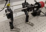
PUMA is an advanced direct vision 3D printed multimodality microscopy system with fluorescence ,phase contrast, Köhler illumination and augmented reality functions - and more.

Temperature control in light microscopy – challenges and solutions

Sponsored by Interherence, on 9 September 2021

We describe common challenges that researchers face when trying to precisely control the temperature of the sample they are imaging. We aim to show that thermal control in high sensitivity microscopy is not trivial if you care about temperature directly in the field of view of the microscope (which you should). We hope to persuade

Tissue clearing: what invisible samples reveal about biology

Posted by , on 8 September 2021

Written by Jorge Almagro and Hendrik Messal The tissues that constitute organs exist in our body in 3D. However, for practical reasons, histological analysis has been traditionally performed in 2D, slicing a few micrometer-thick sections to analyse them under the microscope. While this has been critical to understand our anatomy for centuries, it constitutes a simplification of

Dragonfly: The Ideal Confocal System for Expansion Microscopy

Sponsored by Andor, on 30 August 2021

Challenges in Imaging Expanded Samples The resolution in light microscopy is limited by the diffraction limit of light, which means that structures that are closer than 200 nm apart cannot be distinguished unless researchers use Super-resolution techniques. An alternative approach to super-resolution methods was developed by Edward Boyden’s laboratory at MIT in 2015 [1]. Instead of increasing

Turn Your Inverted Microscope into a Multimodal Nanoscope

Sponsored by Olympus, on 14 June 2021

Among recent nanoscopy techniques that break the diffraction limit, single-molecule localization microscopy (SMLM) contributes to major discoveries in medicine and biology. It is now possible to see how subcellular molecular machineries form and behave inside single cells and to quantify single biological molecules such as proteins, nucleic acids, and lipids, at ultralow concentrations inside the

What is Expansion Microscopy?

Sponsored by Andor, on 27 May 2021

How can you get the most information from expanded samples? Traditional light microscopy is limited by the diffraction of light, consequently, features less than 200 nm apart cannot be resolved. For a significant time microscopy technique development was focused towards improving imaging techniques to allow for individual molecules to be resolved. Super-resolution microscopy was developed

Compact and High Performance Fluorescence Microscopy

Sponsored by Etaluma, on 28 April 2021

“Disruptive technologies typically enable new markets to emerge… disruptive products are simpler and cheaper;” Clayton M. Christensen, The Innovator’s Dilemma Microscopes are the most common laboratory instrument in the world; there are more labs with a microscope in them than any other instrument.  As such, it is a field where innovation is constantly producing new

Part V The future: The hope of smart microscopes and phantoms

Posted by , on 24 April 2021

Elisabeth Kugler 1 and Emmanuel G. Reynaud 2 Contact: kugler.elisabeth@gmail.com; emmanuel.reynaud@ucd.ie We are reaching the end of our LSFM journey. Now it is time to look into the future, riding the plane of light! So, what is next for the LSFM? 1. Four-to-two, then four, then one. The story of SPIM, and LSFM per se,

Technology highlights - Quantitative Phase Imaging (QPI)

Posted by , on 20 January 2021

Interview with Helena Chmelová, Ph.D. from the Light Microscopy Core facility at the Institute of Molecular Genetics of the Czech Academy of Sciences in Prague, Czech Republic. Can you tell us a little bit about yourself – where you work, what your research focus is? I work at the Light Microscopy Core Facility at the Institute of

Technology highlights - Traction Force Microscopy (TFM)

Posted by , on 9 December 2020

Interview with Aki Stubb, Ph.D. Please tell us a bit about yourself and the facility where you work. My name is Aki Stubb and I am a post-doctoral fellow at the University of Cambridge, UK. I did my PhD in the group of Johanna Ivaska at the Turku Bioscience Centre, University of Turku and Åbo