Advertisement

Displaying posts in the category: How to

Conference Tips from the yDGE

Posted by , on 26 April 2023

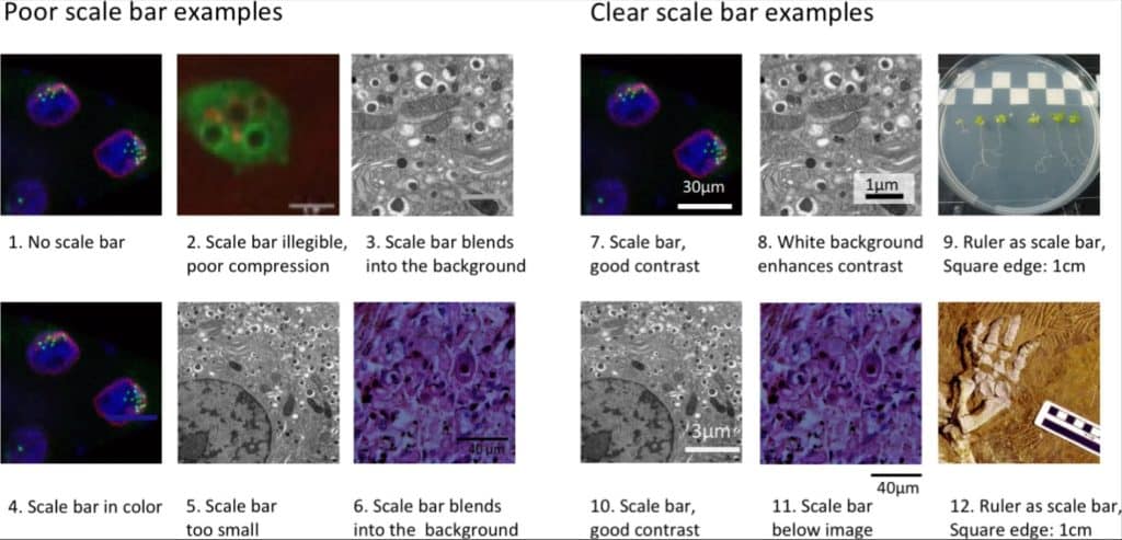
About yDGE DGE Young Microscopists, abbreviated yDGE, is a working group within the German Society for Electron Microscopy (DGE) for students, doctoral researchers, as well as early-career post-doctoral researchers and professionals. In January 2022, we started as a small (but all the more motivated) group of peers. Our vision was, and still is, to promote

Bypassing 164 years of tradition with 'any immersion microscopy'

Posted by , on 24 April 2023

The optical microscope is a classic scientific instrument with a straightforward purpose: to observe objects in more detail than is possible with the naked eye. Many microscope variations exist, from the rudimentary examples of the 17th century, to modern computer controlled systems with sophisticated designs. Despite the variety, most optical microscopes reuse the same physical

Quality assurance of segmentation results

Posted by , on 13 April 2023

This blog post revolves around determining and improving the quality of segmentation results. A common problem is that this step is often omitted and done rather by the appearance of the image segmentation than by actually quantifying it. This blogpost aims to show different ways to achieve this quantification as this leads to reproducibility. Therefore,

Annotating 3D images in napari

Posted by , on 30 March 2023

This blog post revolves around generating ground truth in 3D images for segmentation. Therefore, we will define what ground truth is and how we can generate it using napari in a time-efficient way. We will also learn about difficulties of annotating alone or in groups and address possible solutions for both. Challenges of image segmentation

Fast4DReg - to the rescue of your drifty microscopy data

Posted by , on 10 March 2023

Fast4DReg to the rescue of your 4D microscopy data In life sciences, researchers use microscopes to study living organisms, such as cells or small animals. These live cell imaging experiments are usually performed over several hours, exposing the experiment to changes in the sample and in the microscope surroundings – causing the data to drift.

Rescaling images and pixel (an)isotropy

Posted by , on 2 March 2023

This blog post shows the importance of rescaling 3D image data and what anisotropy of pixels/voxels has to do with it. We will see which different options exist to rescale an image as well as their advantages and limitations. We will also apply a commonly used segmentation algorithm, Voronoi-Otsu-Labeling, on the original and the rescaled

Sharing research data with Zenodo

Posted by , on 15 February 2023

TL;DR: Sharing data open access is good scientific practice. If data is shared via online portals such as https://zenodo.org, we can implement best practices for sharing, licensing, reusing and citing research data. In this blog post I guide through the minimal procedures that are necessary to share a dataset publicly following the FAIR principles; to

Managing Scientific Python environments using Conda, Mamba and friends

Posted by , on 8 December 2022

TL:DR: This blog post gives short instructions and explanations to demystify how some scientific Python programmers, including myself, organize their coding environment for the sake of reproducibility of science. We will see what conda and mamba are, what they are good for, and how to use them properly. We will also take a short excursion

60 years of Fluorescent Proteins

Posted by , on 4 October 2022

In talks about fluorescent proteins I usually include a timeline of events related to Green Fluorescent Protein (GFP). The timeline highlights some of the key moments in the history of fluorescent protein discovery and engineering. I am generally fond of timelines, since they provide a way to pay tribute to the pioneers, and other researchers

First steps for presentation and analysis of calcium imaging data

Posted by , on 6 September 2022

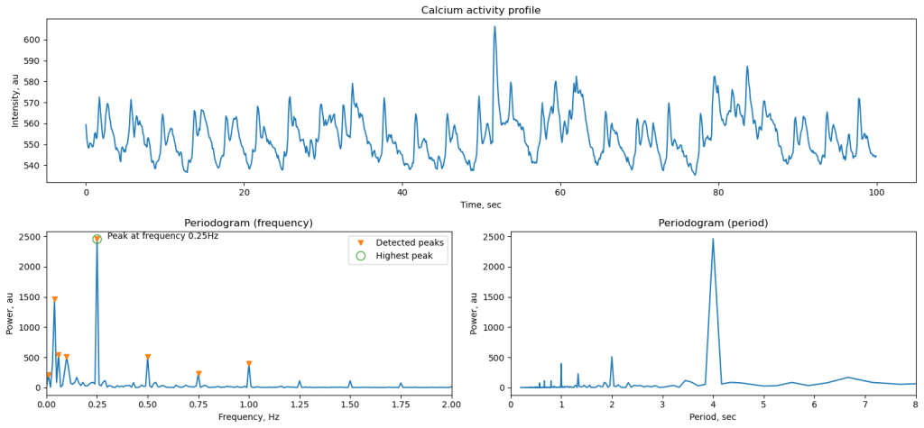
Andrey Andreev, Desiderio AscencioCalifornia Institute of Technology, David Prober labaandreev@caltech.edu Calcium imaging is a widely used tool in neuroscience, and our community now has access to multiple computational tools to process and analyze these data. However, a significant portion of work with imaging data relies on much simpler approaches than offered in these software packages.