/* */

Advertisement

Displaying posts in the category: How to

How to use Dragonfly Spinning Disk Microscope for Multiplex In Situ Hybridization

Sponsored by Andor, on 4 November 2020

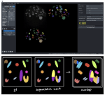
Multiplexing in cell biology is the unveiling of several (Xn) RNAs in its 2D or 3D biological context. Multiplexing has become a hot topic in neurosciences, oncobiology, disease target diagnostics, development, behavioural studies, etc. Several techniques have been developed to allow multiplex imaging; examples of such are FISSEQ, instaSEQ, osmFISH, STARmap, MERFISH and seqFISH. The advantage of

Primers on Microscopy for Biologists - Resolution

Posted by , on 15 July 2020

Formal definitions of resolution refer to imaginary objects such as infinitely small sources of light. I will avoid those and instead try to provide a pragmatic explanation. Practically, the spatial resolution is the size of the smallest structure that can be distinguished in the light coming from a specimen. All sensors and components of the

How CLIJ2 can make your bio-image analysis workflows incredibly fast

Posted by , on 14 July 2020

Do you also spend a substantial amount of your time waiting for automated image analysis to finish? I did, again and again for more than a decade. And, then,  the morning after running a script overnight, you realize that a parameter was wrongly set. It was about time to change that. History Processing on graphics

Fixation artifacts and how to minimize them

Posted by , on 7 July 2020

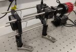
Sample preparation is the first step for having high-quality images that will impress everyone, but it is often overlooked. Many times I have tried to help others improve their microscope images, only to find out that improvement was not possible due to the quality of the sample. No matter how expensive your microscope is, you

The cell biologist's guide to super-resolution microscopy

Posted by , on 1 July 2020

Super-resolution microscopy has allowed to resolve cellular structures down to the nanoscale. While extremely beneficial for most cell biologists, the advent of this technology has made it somewhat difficult for cell biologists and microscopists alike to select the techniques best suited for their experiments. Guillaume Jacquemet, Alexandre Carisey, Hellyh Hamidi and two members of our