Advertisement

Displaying posts with the tag: is_archive

Workshop report: France BioImaging - Advanced Training on Light-Sheet Microscopy

Posted by , on 22 May 2025

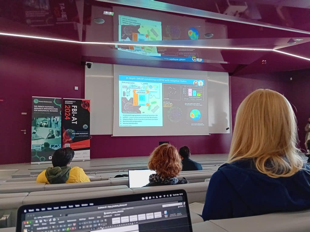
From November 4th to 7th, 2024, I had the privilege to attend the Advanced Training on Light-Sheet Microscopy‘s Principles and Applications to Neuroscience and 3D Cell Culture at France BioImaging in Bordeaux. This comprehensive training, rich in both theoretical and practical knowledge, provided an exceptional platform for learning and engaging with experts in the field.

Enhancing Global Access: interview with CZI grantee Alenka Lovy

Posted by , on 24 January 2024

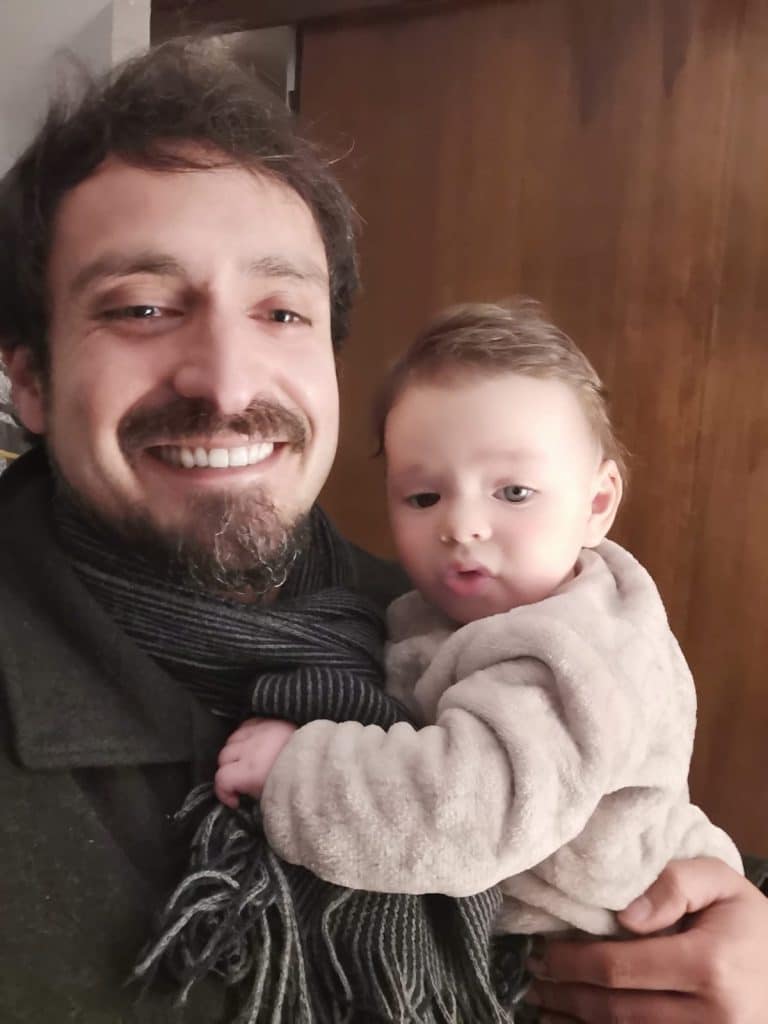
DEVELOPING A LATIN AMERICAN LIGHT-SHEET BIOIMAGING HUB Alenka Lovy is a principal investigator at Universidad Mayor in Chile. She is also the leader of LiSIUM (the Light Sheet Imaging Unit at Universidad Mayor). Her CZI project aims to connect multiple labs in the region, including institutions in Chile, Brazil, and Uruguay, to implement light sheet

Enhancing Global Access: interview with CZI grantee Michael Weber

Posted by , on 24 January 2024

UNLOCKING NEW BIOLOGICAL RESEARCH WITH MODULAR AND SHAREABLE ‘FLAMINGO’ LIGHT SHEET MICROSCOPES Michael Weber has focused a lot of his career in microscopy development. He is a grantee of the CZI cycle 1 grants to imaging scientists. As part of his project, he helps developing light sheet microscopes called ‘Flamingos’ that can travel between research

An interview with Alenka Lovy

Posted by , on 27 November 2022

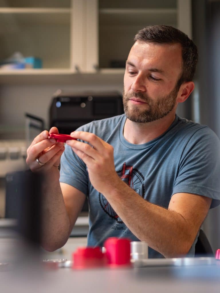
MiniBio: Dr. Alenka Lovy is an Assistant Professor at the Center for Integrative Biology (CIB) and in charge of LiSIUM, LightSheet Imaging at Universidad Mayor. where together with a team of researchers, she is introducing this novel platform to the Latin American region. Alenka is originally from the Czech Republic. She did her early studies

An interview with Ramón Ramírez

Posted by , on 8 November 2022

MiniBio: Dr. Ramon Ramirez is the Coordinator of the Microscopy Centre at Universidad Mayor in Santiago de Chile, and a leading member at LiSIUM (the light sheet imaging centre). He did his early career in Chile, at Pontificia Universidad Católica de Valparaíso where he studied a BSc in Biology. He later went to Madrid to

Tissue clearing: what invisible samples reveal about biology

Posted by , on 8 September 2021

Written by Jorge Almagro and Hendrik Messal The tissues that constitute organs exist in our body in 3D. However, for practical reasons, histological analysis has been traditionally performed in 2D, slicing a few micrometer-thick sections to analyse them under the microscope. While this has been critical to understand our anatomy for centuries, it constitutes a simplification of

FocalPlane features... Pavel Tomancak

Posted by , on 7 June 2021

In case you missed ‘FocalPlane features…’ this month, here is the recording of the webinar given by Pavel Tomancak on Tuesday 1 June 2021. Light sheet microscopy has been over more than a decade producing stunning new views of the dynamics of living systems. I have been observing the rapid growth of this technology, contributed

Part V The future: The hope of smart microscopes and phantoms

Posted by , on 24 April 2021

Elisabeth Kugler 1 and Emmanuel G. Reynaud 2 Contact: kugler.elisabeth@gmail.com; emmanuel.reynaud@ucd.ie We are reaching the end of our LSFM journey. Now it is time to look into the future, riding the plane of light! So, what is next for the LSFM? 1. Four-to-two, then four, then one. The story of SPIM, and LSFM per se,

LSFM series – Part IV: Data post-processing: Cropping, tailoring, and trimming

Posted by , on 23 January 2021

Elisabeth Kugler 1 and Emmanuel G. Reynaud 2 Contact:kugler.elisabeth@gmail.com; emmanuel.reynaud@ucd.ie There is a very thin sheet of light between gathering data and hoarding. In Science, between pilling up manuscripts on desk, books on shelves and samples in cold freezer, most of us fit in the latter category. It is OK, if you have space available

LSFM series – Part III: Image acquisition: Calibration and acquisition

Posted by , on 17 December 2020

Elisabeth Kugler 1 and Emmanuel G. Reynaud 2 Contact:kugler.elisabeth@gmail.com; emmanuel.reynaud@ucd.ie Now, we are getting closer to the point of setting up our sample in the chamber to image it. Now that we know which microscope we are about to use (Part I) and have mounted it the right way (Part II), we need to trust