Advertisement

Displaying posts in the category: Default

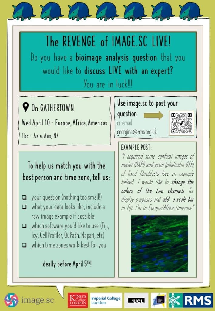
The Revenge of Image.sc LIVE! around the world - the live bioimage analysis helpdesk is returning to a time zone near you!

Posted by , on 25 March 2024

Community surveys often point to the biggest bottleneck in excellent bioimaging science being image analysis. That’s why the RMS DAIM committee and their friends across the world are putting on another event to highlight the fantastic image.sc forum. All the info you need is here: Image.sc LIVE around the world! – Announcements – Image.sc Forum and in the poster

Image Analysis course with Fiji/ImageJ

Posted by , on 20 March 2024

An online course introducing you to the basics of image analysis, including automatic segmentation, colocalisation, denoising, 3D, etc. Two sessions, happening in May and June 2024.

Will your algorithm be the best for this new image data challenge?

Posted by , on 26 February 2024

In order to answer image data analysis demands, France-BioImaging is launching its first data machine learning competition: welcome to the Light My Cells challenge! The Light My Cells challenge aims at contributing to the development of new image-to-image ‘deep-label’ methods in the fields of biology and microscopy. Basically, the goal is to predict the best-focused output-images of several organelles

Image Analysis course with Fiji/ImageJ

Posted by , on 20 February 2024

An online course introducing you to the basics of image analysis, including automatic segmentation, colocalisation, denoising, 3D, etc.

Enhancing Global Access: interview with CZI grantee Mahmoud Maina

Posted by , on 24 January 2024

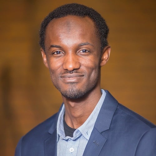
BIOIMAGING NETWORK IN WEST AFRICA Mahmoud Maina is an Associate Professor at Yobe State University in Nigeria and Independent Research Fellow at the University of Sussex. He is the founder and Director of the biomedical Science Research and training Centre (BioRTC) at Yobe State University. His project aims to establish a West African Bioimaging Network

Image.sc LIVE! around the world - a live bioimage analysis helpdesk is coming to a time zone near you!

Posted by , on 13 December 2023

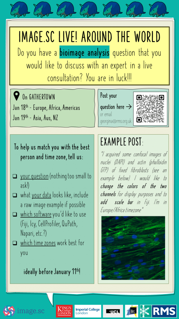
Community surveys often point to the biggest bottleneck in excellent bioimaging science being image analysis. That’s why the RMS DAIM committee and their friends across the world are putting the fantastic image.sc forum “on the road” and staging a first of its kind event which we’re calling “Image.sc LIVE! around the world”. All the info you need is here: Image.sc LIVE

Image Analysis course with Fiji/ImageJ

Posted by , on 3 December 2023

We would like to invite you to an online course introducing you to the basics of image analysis, including automatic segmentation, colocalisation, denoising, 3D, etc. This course will start on February 5th, 2024. Sign-up and additional details here. What You’ll Learn This course consists of five 2.5-hour live interactive online sessions, where you’ll dive into

RMS Application Coaching and Personal Mentoring Scheme

Posted by , on 2 August 2023

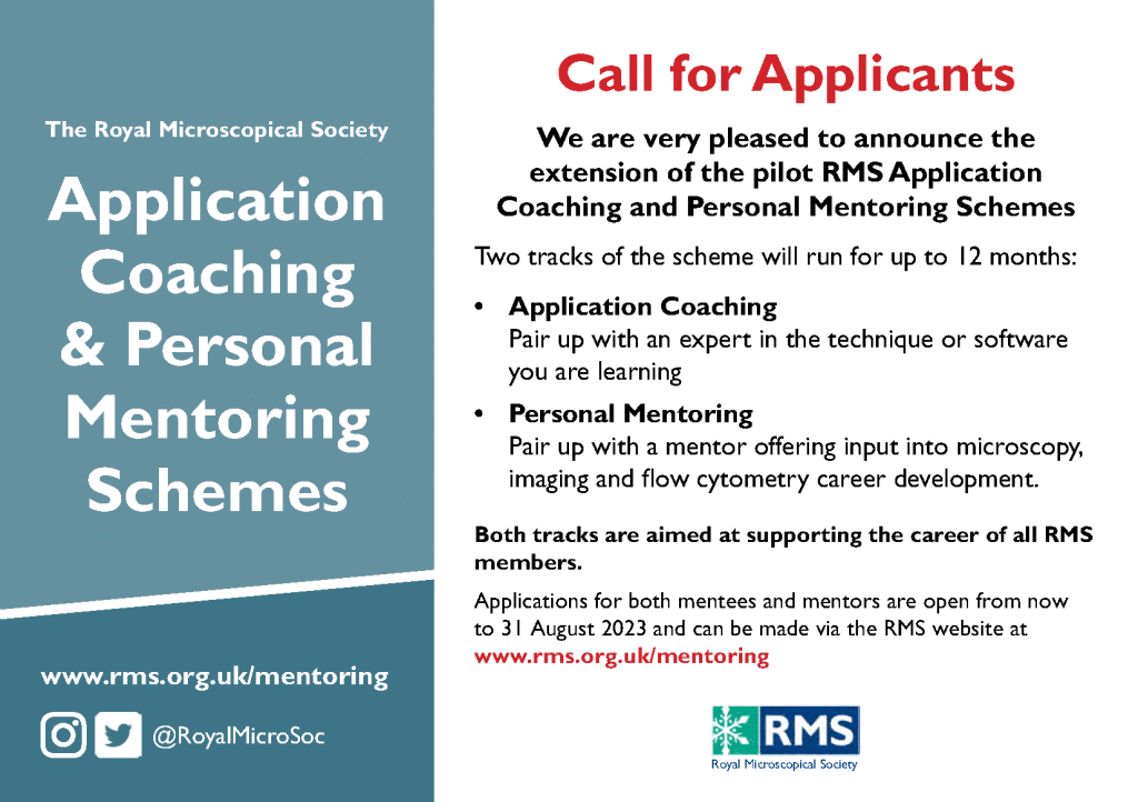
The RMS Application Coaching and Personal Mentoring Scheme pilot was extended to a twice yearly call for applicants. We are accepting applications until the 31 August 2023 with the next round opening in January 2024. For more details and to apply as a Mentor, Mentee or both please visit this webpage: https://www.rms.org.uk/opportunities/mentoring-schemes.html

NEW Technical Specialist Job Shadowing Scheme!

Posted by , on 2 August 2023

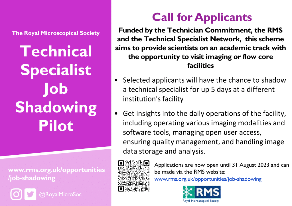
The Technical Specialist Job Shadowing Scheme is a brand new initiative supported by the RMS, Technician Commitment and Technical Specialist Network that is designed to provide scientists on an academic track with an opportunity to visit a UK imaging or flow core facility for up to 5 days. Applications close on 31 August. To find

ASCB Subgroup: Deep Learning and Artificial Intelligence in Cell Imaging

Posted by , on 17 July 2023

The deep learning revolution in bioimage analysis over the past ~5 years has been nothing short of awe-inspiring; in just a few short years we’ve gone from having very few deep learning tools usable on light microscopy images to new approaches coming out weekly and routinely outperforming older classical approaches. We can now use deep