/* */

Advertisement

Featured image with Martín Estermann

Posted by , on 15 September 2023

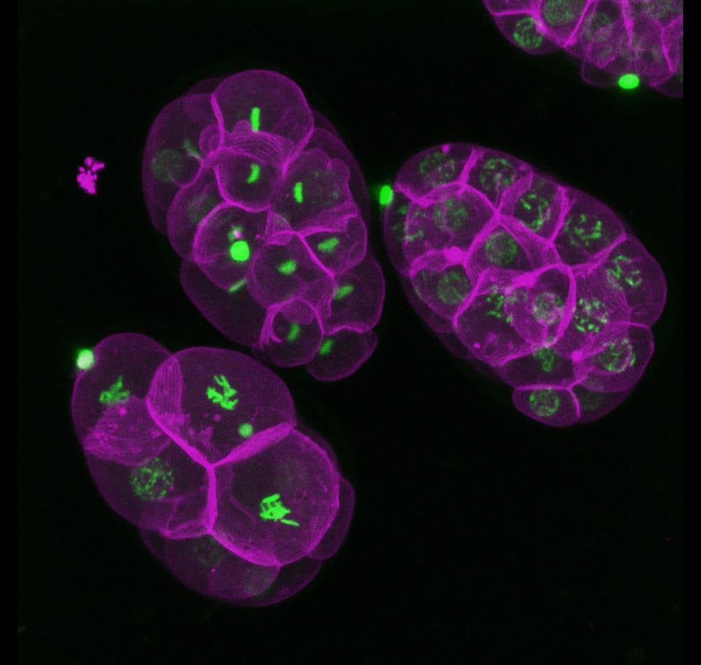
Our featured image, acquired by Martín Estermann, is a 3D reconstruction of C. elegans transgenic embryos. Embryos were imaged live using a Zeiss Celldiscoverer 7 microscope. The 3D reconstruction was performed using Fiji. Histones in are shown in green and the cell membrane in magenta. We caught up with Martín to find out about his

'How they started with Euro-BioImaging & Global BioImaging (GBI) ?'

Posted by , on 14 September 2023

As a continuation to the series of blogs called “how it started…”, where we shed light on the journey through the origins of various imaging communities around the world, we speak to Euro-BioImaging and GBI. Our mission for this series is simple yet powerful – by learning about the visions and goals of these communities,

Working smarter with LEDs and optical filters for widefield fluorescence

Sponsored by CoolLED, on 13 September 2023

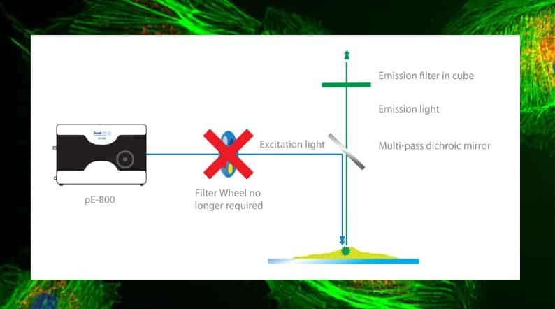
Optical filters can make or break a fluorescence microscopy experiment. For widefield fluorescence, they also have the potential to unlock breakthroughs in high-speed, high-contrast imaging when used alongside multi-channel LED illumination systems. There are thousands of filters and dichroic mirrors to choose from, with the selection depending on the fluorophore(s) and light source. While imaging

An interview with Manuel Forero

Posted by , on 12 September 2023

MiniBio: Prof. Manuel Forero is a professor and principal investigator at Universidad de Ibagué, in Colombia, where he focuses on pattern recognition, and the development of image processing and machine learning techniques for biomedical and microscopy applications. He graduated as an Electrical Engineer from Universidad Javeriana, in Bogotá, Colombia. He went on to pursue MSc

20 years of the York confocal course

Posted by , on 11 September 2023

The confocal course at the University of York has been running for 20 years, and I caught up with newly elected RMS President Peter O’Toole at #mmc2023 to discover the history of the course and to find out why it is so successful. With many alumni from the course at the meeting, including at the

PoLBIAS23 - Event Reflections and Revelations

Posted by , on 8 September 2023

Reflections and Revelations: Highlights from #PoLBIAS23 - A Symposium on Bioimage Analysis. This blog showcases a summary, as well as a reflection on open questions and community efforts,.

Microscopy preprints – new tools and techniques in imaging

Posted by , on 8 September 2023

Here is a curated selection of preprints posted recently on new tools and techniques in imaging. Let us know if we are missing any preprints that are on your reading list.

Imaging with the Liverpool Centre for Cell Imaging (CCI)

Posted by , on 7 September 2023

In the first of our series of 'Imaging with...' blog posts, we meet the staff at the Liverpool CCI

A schedule for organizing international symposia

Posted by , on 5 September 2023

TL;DR: Coordinating an international conference can be a complex endeavor, especially for scientists whose primary focus often lies outside the realm of event management. A major challenge in this context is timing. In this blog post I outline a potential schedule of tasks for organizing an international conference such as the PoLBIAS23. As a senior

All things unequal – live microscopy reveals asymmetric RNA localization in the early mammalian embryo

Posted by , on 4 September 2023

Authors: Jennifer Zenker1,*, Julian Heng2 1Australian Regenerative Medicine Institute (ARMI), Faculty of Medicine, Nursing and Health Sciences, Monash University, Clayton, Australia 2Remotely Consulting, Perth, Australia *Corresponding author Non-mammalian systems have been invaluable for us to decipher evolutionarily-conserved processes, orthologous molecules and cellular phenomena underlying early embryo development. One fundamental discovery from the work on non-mammalian