Advertisement

Displaying posts with the tag: is_archive

PoLBIAS23 - Event Reflections and Revelations

Posted by , on 8 September 2023

Reflections and Revelations: Highlights from #PoLBIAS23 - A Symposium on Bioimage Analysis. This blog showcases a summary, as well as a reflection on open questions and community efforts,.

Tracking in napari

Posted by , on 1 June 2023

In this blog post we are exploring cell tracking in napari. Two important processes in normal tissue development and disease are cell migration and proliferation. To gain a better understanding on these processes, tracking in time-lapse datasets is needed. By measuring track properties, like velocity and the total travelled distance, spatio-temporal relationships can be studied.

An interview with José Luis Vázquez Noguera

Posted by , on 7 February 2023

MiniBio: Dr. Jose Luis Vazquez is a researcher at Facultad Politecnica at Universidad Nacional de Asuncion in Paraguay. Between 2004 and 2008, he studied his undergraduate degree in Computer Systems Engineering at Instituto Tecnológico de León in México as an SRE scholar. Between 2009 and 2012 he completed his MSc degree as an Itaipú Binacional

An interview with Horacio Legal

Posted by , on 31 January 2023

MiniBio: Prof. Horacio Legal is a group leader in the area of Image Analysis at Universidad Nacional de Asuncion in Paraguay. He is also the co-founder of the postgraduate program in the area of Computer Science at this university. Horacio completed his Bachelors degree in Electrical Engineering at the Universidade Federal do Paraná, in Brazil in

First steps for presentation and analysis of calcium imaging data

Posted by , on 6 September 2022

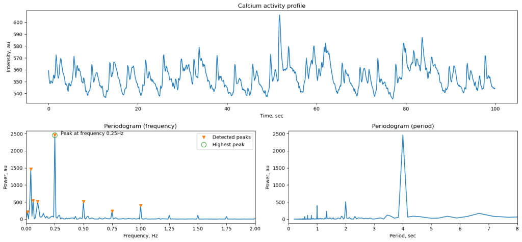
Andrey Andreev, Desiderio AscencioCalifornia Institute of Technology, David Prober labaandreev@caltech.edu Calcium imaging is a widely used tool in neuroscience, and our community now has access to multiple computational tools to process and analyze these data. However, a significant portion of work with imaging data relies on much simpler approaches than offered in these software packages.

An interview with Federico Lecumberry

Posted by , on 5 July 2022

MiniBio: Federico Lecumberry is an Associate Professor in Signal Processing and Machine Learning at the Department of Electrical Engineering at the Faculty of Engineering of Universidad de la Republica in Uruguay. He is also an Associate Researcher with the Advanced Bioimaging Unit of Institut Pasteur de Montevideo. He completed his BSc, MSc and PhD degrees

#GliaMorph - a tool to analyse glial cell morphology in 3D

Posted by , on 17 May 2022

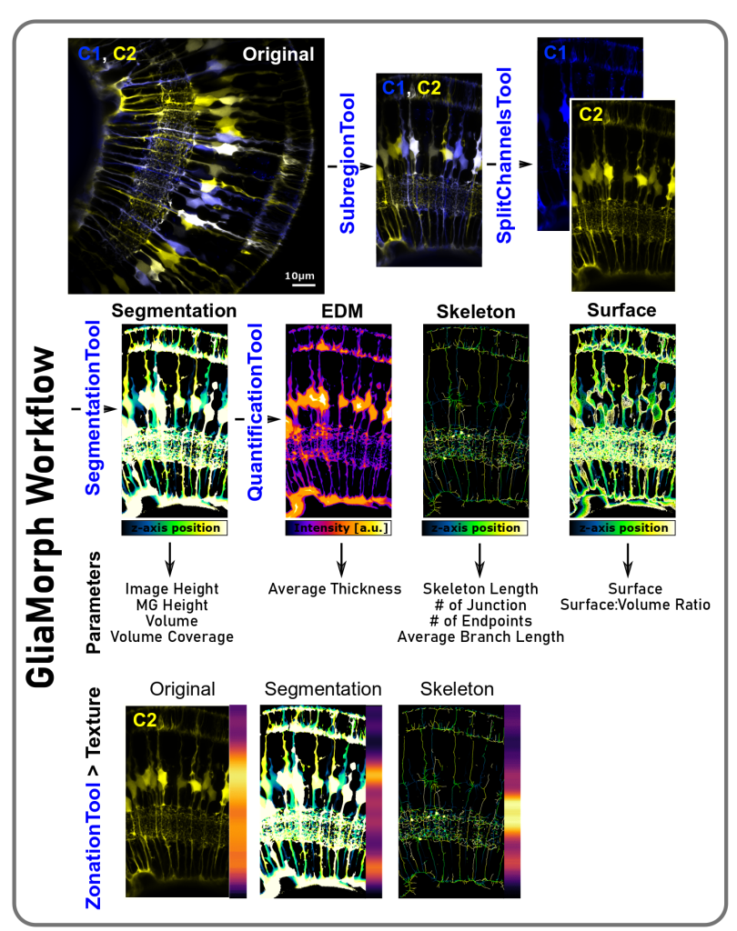
Contents What is #GliaMorph? What can you do and analyse with #GliaMorph? Links to manuscripts and data Contributions and Feedback Thanks to all contributors and people providing feedback References What is GliaMorph? GliaMorph is a toolkit implemented as Fiji Macros to allow modular application to process and analyse Müller glia morphology in the retina. During

Job Opportunity: Light Microscopy and Image analysis at the Centre for Cellular Imaging of Gothenburg University

Posted by , on 4 February 2022

We are looking for a new colleague to join the light microscopy team of the Centre for Cellular Imaging of Gothenburg University (CCI). Information below and in this link Researcher, Core Facilities The University of Gothenburg tackles society’s challenges with diverse knowledge. 53 500 students and 6 500 employees make the university a large and inspiring place

Janelia+EMBL BioImaging Seminar Series: How We Started a Successful Seminar Series during the Pandemic

Posted by , on 25 January 2022

How we started a global BioImaging seminar series in the middle of a global pandemic...

DeepImageJ: Deep learning in bioimage analysis for dummies

Posted by , on 2 December 2021

Written by Estibaliz Gómez-de-Mariscal, Cristina de-la-Torre-Gutiérrez, Arrate Muñoz-Barrutia, and Daniel Sage Do you invest significant time looking for tools to analyze your microscopy images? Probably you have already heard about the extraordinary capabilities of deep learning (DL) to reveal complex visual structures in images. If you are a biologist practicing daily ImageJ, you may look